
Third eyelid prolapse in dogs
Many vertebrates have a third eyelid—a small fold of conjunctiva—at the inner corner of their eyes. This membrane protects and cleanses the eyeball during blinking, while the lacrimal gland located beneath it moistens the cornea, preventing it from drying out. In some cases, the lacrimal gland enlarges and shifts from under the eyelid outward. This pathological condition, called third eyelid prolapse, can occur in dogs as a result of certain diseases or due to a breed predisposition.
Content
Causes of the development of pathology
Third eyelid prolapse in dogs can be caused by:
- corneal injury or foreign body lodged under the eyelid;
- dislocation or subluxation of the lens;
- eversion of the eyelid (ectropion);
- adenoma of the 3rd eyelid (anatomical defect of the nictitating membrane);
- trichiasis (direction of eyelash growth towards the cornea of the eye);
- dry keratoconjunctivitis;
- acute glaucoma (increased intraocular pressure).
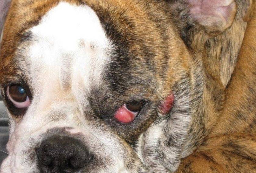
Prolapse of the lacrimal gland of the third eyelid is diagnosed more frequently in dogs than in cats and typically develops in puppies under one year of age. Typically, a few months after the prolapse appears in one eye, the other eye is also affected. Breeds prone to this condition include Mastiffs, Bulldogs, Shar Peis, Pugs, Pekingese, Cane Corsos, Chihuahuas, and Toy Terriers. Experts attribute this genetic predisposition to hyperplasia of the lacrimal gland and weakness of its muscular ligament in these breeds.
In cats, prolapse is mainly found in representatives of brachycephalic breeds with a wide skull and a flattened muzzle - these are Persian, Scottish Fold, British, and Himalayan.
Symptoms and diagnosis
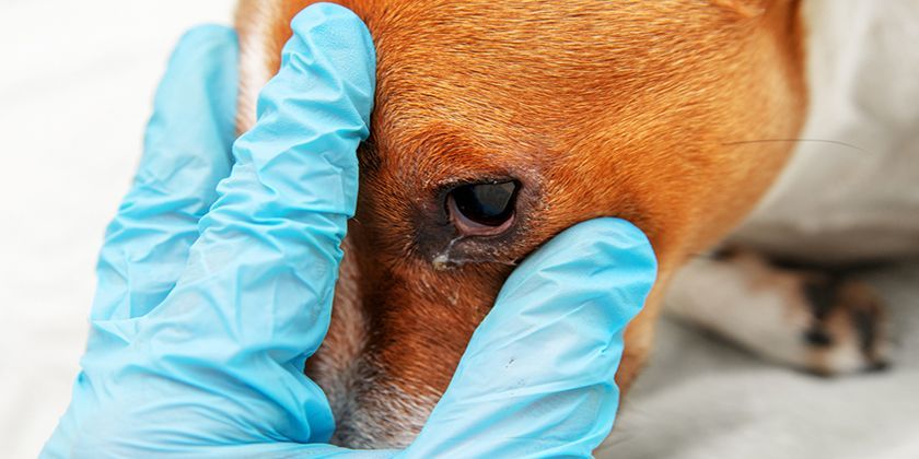
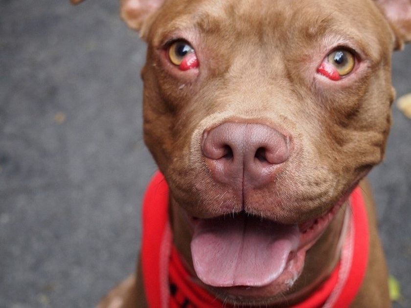
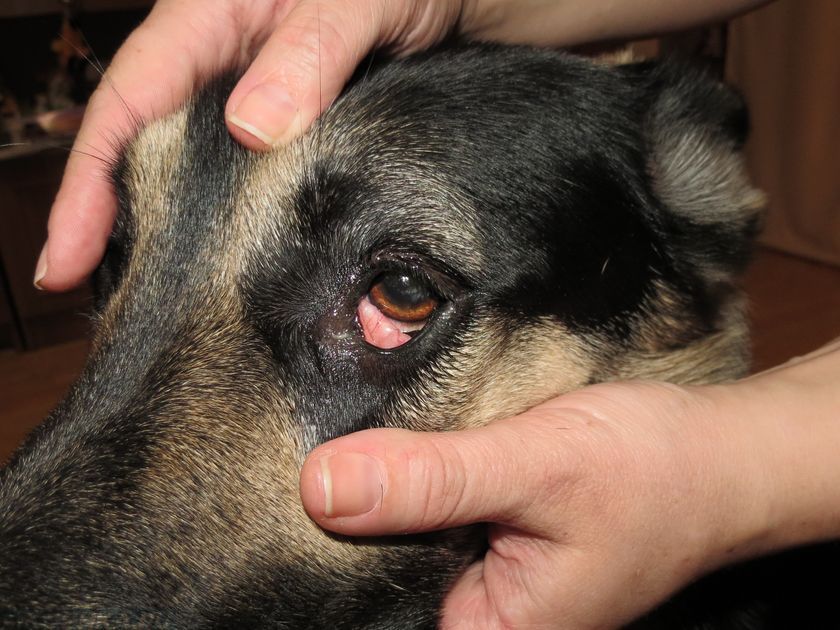
In its initial stages, third eyelid prolapse in dogs manifests as a small, round, pink swelling that periodically appears in the corner of the eye. Over time, the displaced gland becomes pinched, swells, and turns dark red. Redness of the entire conjunctiva, profuse lacrimation, and blepharospasm (frequent involuntary contractions of the eyelid muscles) are observed. The eye becomes purulent, and the dog constantly tries to scratch it.
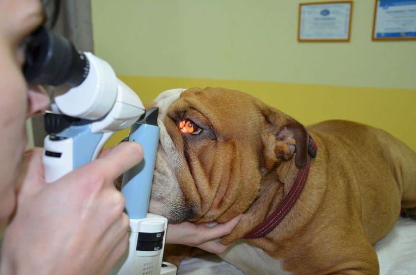
A preliminary diagnosis is made based on a visual examination by a veterinary ophthalmologist. It is important to differentiate lacrimal gland prolapse in dogs from other pathologies, particularly third eyelid hypertrophy caused by inflammation. Therefore, if necessary, diagnostic procedures may include:
- examination of the third eyelid with tweezers under local anesthesia;
- measurement of intraocular pressure (redness of the whites of the eyes may be caused by ocular hypertension);
- testing of pupils (the cause of conjunctival hyperemia may be the inflammatory disease uveitis);
- biomicroscopy of the anterior wall of the eye (examination using a special lamp with a microscope, which allows one to detect corneal damage or a foreign body getting on it).
- neurological examination (a number of neurological diseases can lead to decreased muscle tone of the eyelid or protrusion of the eyelid).
- Ultrasound of the eye and tissues of the eye orbit, X-ray of the skull (to detect possible anatomical pathologies).
Treatment
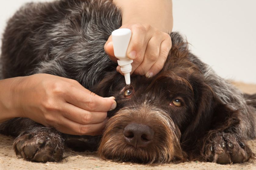
Treatment for third eyelid gland prolapse requires surgical intervention. In the early stages of the condition, symptoms of inflammation, irritation, and tearing can be relieved with Keratostil, Burdi, Multi Avizor Moisture, and Nutri Vet Eye Cleanse eye drops. However, these will not resolve the problem, as they will not return the prolapsed gland to its proper place.
The purpose of the surgical procedure depends on the cause of the pathology: if it is a rupture or weakening of the ligament, the lacrimal gland is not removed, but rather fixed in the correct position. Resection of the gland is only necessary if there is a tumor on it, since without the lacrimal gland, the eyeball will be deprived of sufficient lubrication, which can lead to the development of pathologies such as dry conjunctivitis or keratitis.
A week before the surgery, the dog undergoes a course of antibiotic therapy. The surgery is performed in the clinic under general intravenous anesthesia using a binocular loupe. There are two options for fixing the gland in the anatomically correct position.
Pocket technique
The method involves placing the prolapsed gland into a pocket created by making small conjunctival incisions on both sides of the gland. These incisions are then sutured with thin absorbable suture. This suture is gentle on the animal, does not injure the cornea, and is guaranteed to prevent recurrent prolapse.
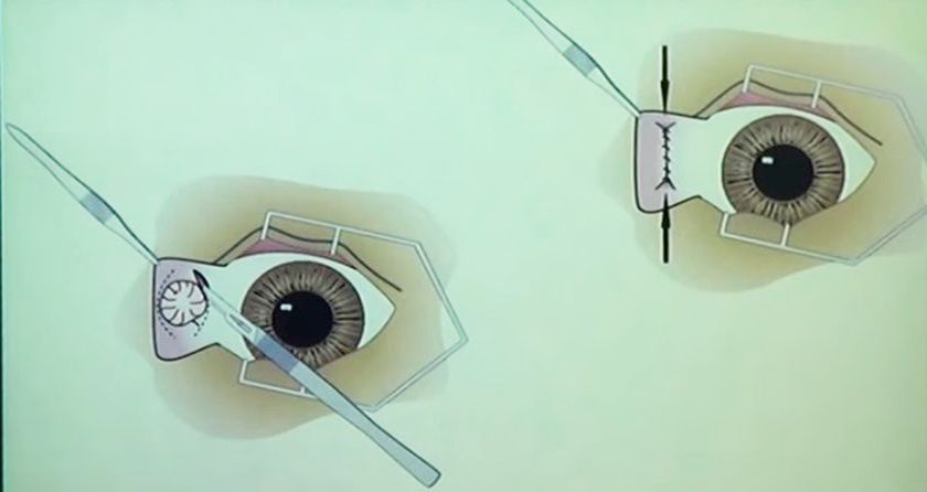
Purse-string suture technique
This method of prolapse repair involves placing a ring-shaped suture around the gland on the bulbar surface of the third eyelid. When the suture is tightened, the gland moves under the eyelid and assumes its correct position. The suture knot is placed on the palpebral (eyelid-facing) side of the conjunctiva, using absorbable suture material.
Rehabilitation period
After surgery, the dog is prescribed antibiotics, nonsteroidal anti-inflammatory drugs, and immunomodulatory medications for one to two weeks. To prevent injury to the eye, the dog is placed in a protective collar until the sutures are completely healed.
With timely treatment, third eyelid prolapse in dogs has a favorable prognosis: statistically, complete recovery occurs in 95-98% of cases. Follow-up examinations using a slit lamp and assessing the gland's tear production using the Schirmer test are typically performed by the veterinarian two weeks and two months after surgery.
Read also:


Add a comment