
Lipoma in dogs: what does it look like and how to remove it
A lipoma in dogs is a benign tumor in the form of a mobile subcutaneous lump that forms as a result of the proliferation of fat cells. The medical term for such growths is lipoma. If poorly cared for and not treated promptly, they can significantly increase in number, appearing throughout the body or congregating in one area. This condition is called lipomatosis. Lipomas in dogs can form on internal organs, but most often they develop under the skin, causing no discomfort unless they are located in areas prone to injury or simply become a nuisance. Ignoring them is essential under any circumstances, as there is a risk of infiltration, cancerous cell degeneration, and other dangerous complications.
Content
Description and types of lipomas
The tumor is a capsule filled with a mixture of fatty and connective tissue. If the fatty component is predominant, the lump remains soft, while if the connective tissue content predominates, it becomes firmer. Less common are non-encapsulated lipomas, which can spread over a large area of the body.
Depending on the location, such neoplasms are of three types:
- superficial – subcutaneous nodules that do not pose a threat to the animal;
- internal - growths on the organs of the abdominal and chest cavity, the danger of which depends on the size and location;
- intermuscular – diffuse formations that penetrate muscle tissue and cause acute pain.
Most often, lipomas in dogs form and are found under the skin—on the neck, head, torso, paws, and tail. To detect internal and intermuscular tumors, an ultrasound and additional laboratory tests are required.
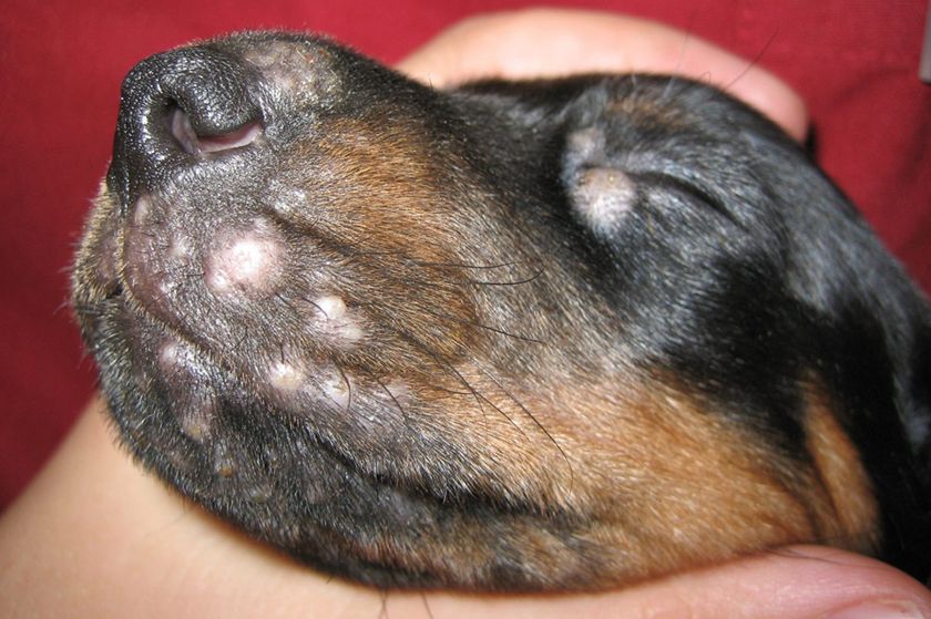
Atheroma, a cyst that develops in a hair follicle due to a blocked sebaceous gland duct, is also often called a lipoma. However, atheroma is unrelated to fatty growths and is not a tumor. To distinguish between these two conditions, it's enough to know what a lipoma looks like on a dog: a photo clearly shows that it has a smooth, uniform surface completely covered with hair, while an atheroma has an opening for the gland's excretory duct. Furthermore, a lipoma can easily move under the skin, while an atheroma is always fused to the skin and often protrudes above the surface, with a different structure.
In the early stages of development, benign subcutaneous lipomas are virtually indistinguishable in appearance from liposarcoma, a malignant tumor that develops from subcutaneous lymphoblasts for reasons that are still unknown.
The main difference between these two neoplasms is the growth rate: lipoma grows slowly, while sarcoma It spreads throughout the animal's body, rapidly increasing in size. Since the initial signs of both tumors are very similar, if you notice small lumps under your pet's skin, you should immediately contact a veterinarian for diagnosis.
Reasons for formation
The main causes of the appearance of lipomas in dogs include the following provoking factors:
- heredity;
- unbalanced diet;
- diseases of the gastrointestinal tract, kidneys, liver;
- incorrect metabolism;
- endocrine disorders;
- unfavorable ecology.
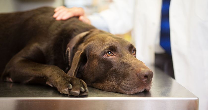
Dogs over 5 years of age are most prone to developing lipomas. Golden Retrievers and Labrador Retrievers are most commonly affected. However, males are less likely to develop these tumors than females.
Possible complications
Superficial growths that are not exposed to any irritants generally pose no threat to the health, life, or well-being of animals. However, there is always a risk of accidental trauma and subsequent malignant transformation. Dogs with a genetic predisposition to cancer or who constantly scratch or bite the lipoma are particularly susceptible to such complications.
Also, the risk of developing complications increases significantly if the lipoma has the following signs:
- dark spots on the surface indicating an increase in melanin concentration due to excessive absorption of UV rays;
- visible growth and increase in size, especially in places where such a growth interferes with movement or compresses blood vessels, nerve fibers and internal organs.
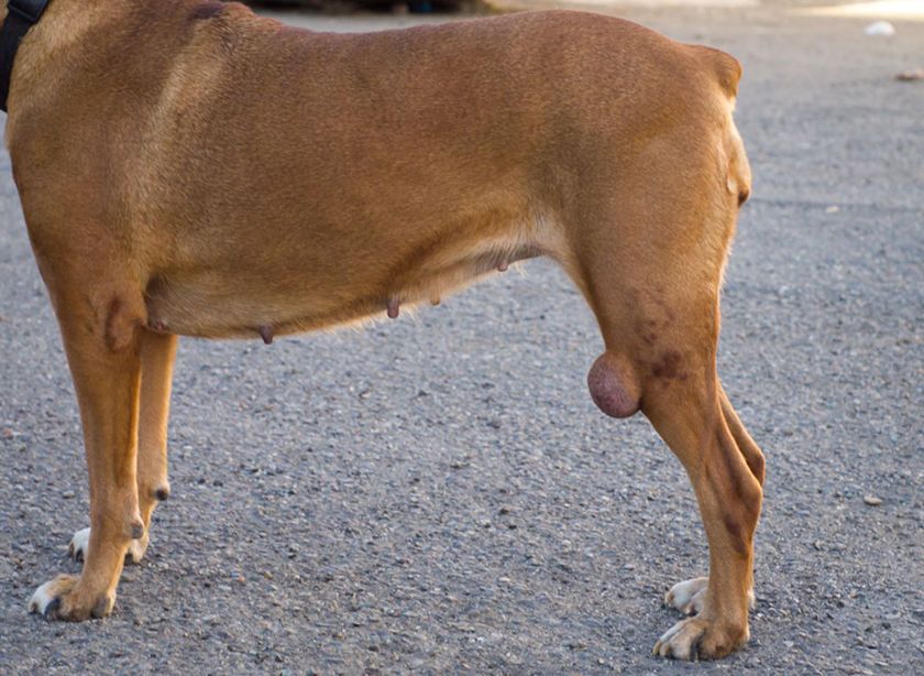
An expanding fatty tumor can change shape, even forming a stalk from which it hangs. This compresses the vessels that supply nutrients and oxygen to the fat cells contained within the capsule. This can lead to tissue death and the gradual development of necrosis, which is extremely life-threatening.
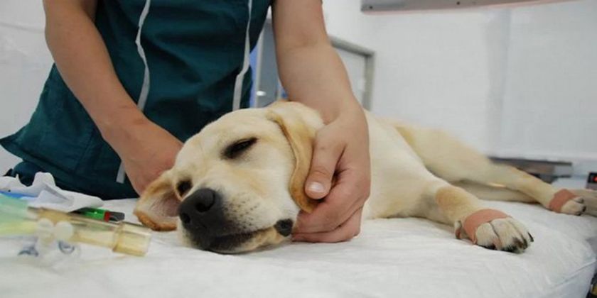
Detection and treatment
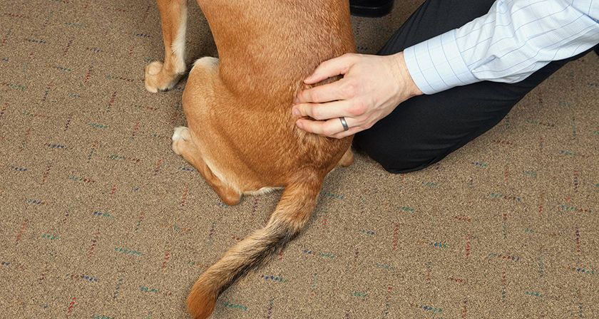
A superficial fatty tumor on a dog's back or other visible areas of the body is often discovered by its owner almost immediately after its appearance. Such growths are especially noticeable in short-haired dogs.
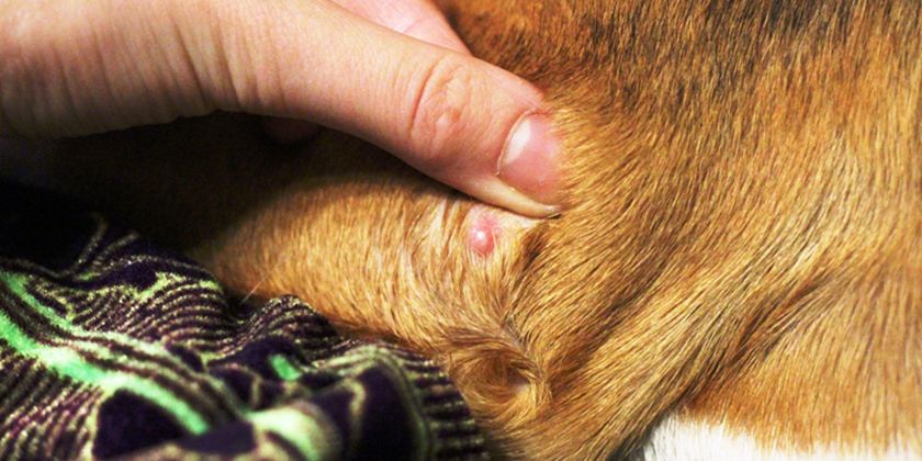
If your pet notices any growths, lumps, or other abnormal changes, they should be carefully examined by parting the fur. If it's a lipoma, it will appear as a small, mobile swelling with a uniform surface. Palpation and pressure should not be painful for the animal.
It's recommended to inspect long-haired breeds during bathing, as their dense undercoat makes a detailed examination significantly more difficult when the coat is dry. It's especially difficult to notice a fatty tumor on a dog's belly, groin, or other hidden area.
If a lipoma is detected, self-medication is not recommended, as any medications or alternative medicine are completely ineffective. Veterinary clinics also do not use medications, physical therapy, or other therapeutic methods to treat this condition, as the fatty and connective tissue that make up the encapsulated lump is indestructible.
The only way to get rid of such tumors and their negative consequences is surgical removal. This is especially important in cases where they swell, grow, or become inflamed.
Veterinary diagnostics
To detect a neoplasm and determine its type, a veterinary clinic uses a comprehensive examination method, which includes the following procedures:
- external examination to determine the size, quantity and location, presence of tissue pigmentation and other characteristics of the tumor;
- palpation of the skin;
- taking a sample of the contents for biopsy.
If the fat capsule is damaged, the tumor is excised with a scalpel. This procedure allows for rapid and complete information about the tumor, as an open wound can become infected. If suppuration is present, surgery is performed urgently.
Internal and intermuscular tumors are detected using ultrasound. Further examinations are prescribed based on the results.
Removal of a lipoma
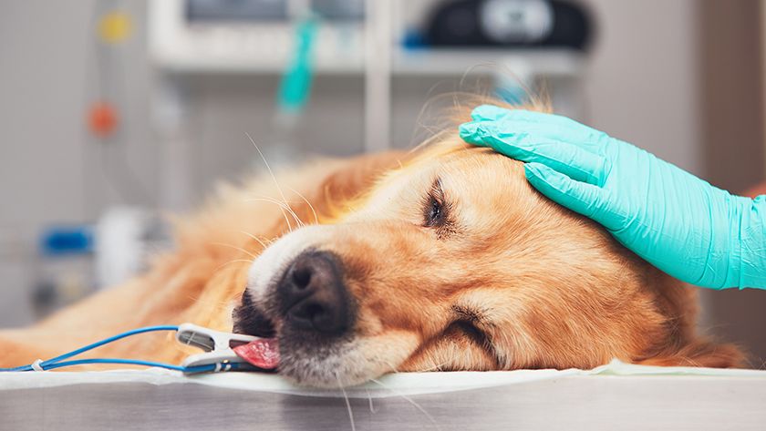
Since a lipoma on a dog's body cannot be treated with conservative methods, it can only be removed through surgery. The surgery is performed in an inpatient setting and is considered straightforward—the pet can be taken home immediately afterward. Small lipomas are excised under local anesthesia, while larger ones are removed under general anesthesiaInternal and intermuscular lipomas, which require layer-by-layer dissection of soft tissue, are removed only under general anesthesia and, as a rule, require a short hospital stay.
The cost of the surgery depends on the size and location of the tumor. In most cases, it ranges from 1,500 to 4,500 rubles. Additional costs may be incurred due to the animal's extended stay at the veterinary clinic.
Before removing a lipoma from a dog, experts recommend following these simple preparatory steps:
- do not feed the animal for 24 hours;
- give only water in sufficient quantity.
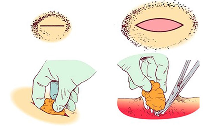
Removing a lipoma in a dog begins with clearing the area of the operation of hair and disinfecting it.
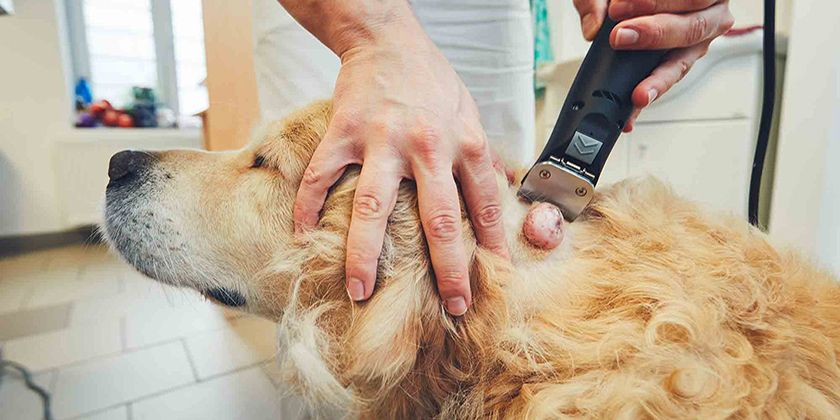
And then everything is done in this order:
- an incision is made over the lipoma;
- the wound is washed to remove any released blood;
- the fat capsule is separated from the vessels and skin;
- the main vessel through which nutrients and oxygen are supplied to the capsule is tied off;
- the capsule is cleared of its contents;
- the cavity is cleared of air and washed with an antiseptic solution;
- the wound is stitched up.
During the recovery period, your pet needs to be provided with proper care, including:
- antiseptic treatment of seams;
- application of antibacterial and anti-inflammatory agents to the wound;
- taking vitamin and mineral complexes and wound healing medications;
- wearing a collar to prevent licking and scratching of the seams.
It's important to note that removal doesn't always completely cure the problem. If the dog is at risk, there's a risk of the tumor recurring. Stress during surgery can also contribute to recurrence.
Therefore, if the tumor is no larger than 1 cm and doesn't bother the animal, it is not removed but kept under constant observation. Such surgeries are also not performed on puppies or older dogs, as they may not tolerate anesthesia well.
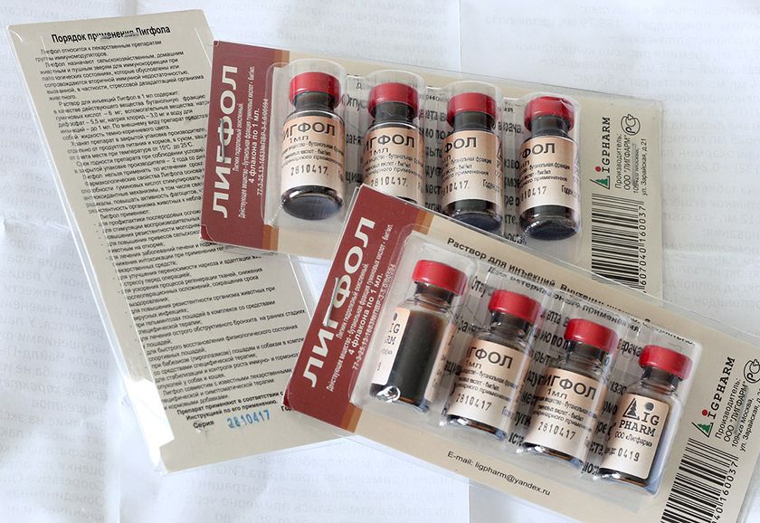
To reduce the rate of fat cell growth and prevent their cancerous transformation, such pets are given Ligfol intramuscularly, with the dosage carefully calculated.
Read also:
- My dog has a lump: what should I do?
- A lump under a dog's skin: causes and treatment
- Tear stains in dogs: what causes them and how to remove them


3 comments
Natalia
We're getting a lipoma removed from our American Cocker Spaniel; she's almost 13.5 years old. It's scary. But the lipoma has burst open and is leaking: blood, pus, and hair stuck to the wound. We're having surgery this week. We're also cauterizing a bunch of other lipomas (there are many small ones all over her body, so we're cauterizing them with nitrogen).
Elena
Did you have surgery? How is the dog?
Olga
A very good article: clearly and understandably written. Thanks to the author! We're planning to remove a lipoma from our toy poodle's armpit (he's almost 10 years old). It doesn't bother him, but it's hanging on a long stalk and is now about 1 cm in size. He's very active, and I'm concerned about damaging the lipoma. I insisted on local anesthesia (the stalk is quite long), but I'm still worried.
Add a comment