
Inflammation of the third eyelid in dogs: symptoms and treatment
Inflammation of the third eyelid is a fairly common condition in dogs. To understand its possible causes and treatment options, it's important to first understand what the third eyelid is and its role in the body.
Content
Forms of pathology
The organ's second name is the semilunar fold. It is located in the inner corner of the eye. One of its main functions is protection from mechanical damage and contamination. In case of danger, the nictitating septum (membrane) instantly closes the eyeball, preventing the entry of foreign objects or softening a forceful blow.
The third eyelid can only be visually seen when a dog blinks. Because the lacrimal glands are located at the base of the semilunar fold, it also serves to lubricate the outer layer of the eye. Lymphoid tissue also serves as an additional barrier against infection.
While this membrane is practically invisible in its normal state, when inflamed, it becomes red, swollen, everted, protrudes outward, and noticeably increases in size. Three main forms of the condition are distinguished:
- Loss.
- Adenoma.
- Volvulus.
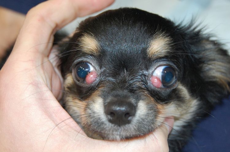
Prolapse of the third eyelid
A fairly common disease in older dogs, as well as breeds with flattened muzzles - French bulldogs, cocker spaniels, Pekingese, Shih Tzu etc. (also found in Cane Corso, Chihuahua) It appears as a prolapse or displacement of the lacrimal gland, appearing as a small reddish-pink swelling in the inner corner of the eye.
The condition can be either permanent or episodic, and the tumor itself can range in size from a few millimeters to several centimeters in diameter. The danger of the disease lies in the fact that when the lacrimal gland is deformed, it stops producing the specific secretion that moistens the eyeball. This, in turn, can lead to the development of associated pathologies, including partial or complete vision loss.
Reasons why the third eyelid droops and falls out:
- Injuries and wounds, foreign bodies (glass, splinters, dust) getting into the eye.
- A sharp increase in intraocular pressure (glaucoma).
- Damage to the lens.
- Decreased contraction function of the muscles located around the eyeball due to physical injury or the development of a number of neurological diseases.
- Consequences of taking tranquilizers and other drugs that affect the nervous system.
- Hereditary predisposition or congenital defects. For example, the presence of abnormally small eyes, which causes the eyeball to "sink" deeper into the orbit, pushing the third eyelid forward.
- Damage to the cranial nerves responsible for contraction of the eye muscles.
- Tetanus. The infection causes convulsions and, as a result, deformation of the third eyelid.
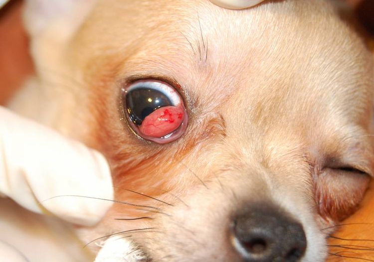
Adenoma
It is a benign tumor that develops in the semilunar fold. It appears as an inverted, pinkish-red growth located in the inner corner of the eye. The condition is relatively rare and is diagnosed by biopsy.
Although the adenoma itself has no effect on the eyeball, it can cause significant discomfort to the animal. The tumor may grow in size or, conversely, disappear temporarily. In the worst-case scenario, its diameter can reach up to 15 mm and be accompanied by catarrhal or follicular conjunctivitis. It's possible that a similar pathology will subsequently develop in the other eye.
The most common treatment for adenoma is complete removal of the third eyelid. This is performed surgically. However, it's important to understand that this type of surgery can lead to eye dysfunction and the development of dry eye syndrome.
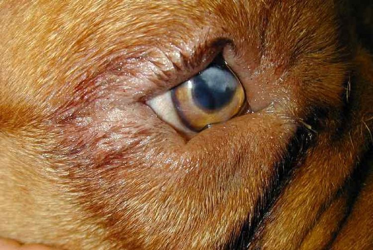
Third eyelid inversion
This phenomenon is typical in young puppies up to 9-10 months of age. The essence of the problem lies in the fact that during the period of active growth, not all organs develop in unison. The ocular cartilage can grow faster than other body parts, and due to its disproportion, it begins to push the third eyelid outward. The anatomical structure of the cartilage changes, leading to swelling and inflammation of the semilunar fold.
Visually, the disease resembles conjunctivitis: the eye swells, turns red, the palpebral fissure narrows, and purulent discharge begins. entropion of the third eyelid Large breed dogs - Dobermans, St. Bernards, Great Danes, German and Central Asian Shepherds, Mastiffs.
Treatment is performed in a hospital setting by excising the deformed portion of cartilage, as seen in the photo below. The surgery is performed under local or general anesthesia using precision optics. It's important to note that any portions of cartilage that have retained their natural shape are not removed.
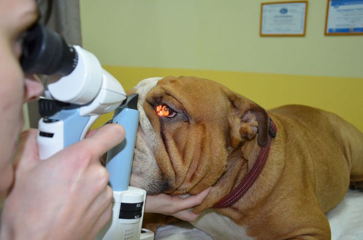
Diagnostics
To collect a complete medical history, the veterinarian interviews the dog's owner and conducts a physical and ophthalmological examination, including pupil testing and intraocular pressure measurement. The following diagnostic tests may also be prescribed:
- Examination of the third eyelid with tweezers under local anesthesia.
- Examination by a neurologist.
- Taking a general blood test.
- Radiographic examination for the presence of a bony orbit.
- Ultrasound of the eye and surrounding tissues inside the orbit.
- Computed tomography of the brain, eyeball, and orbital bone.
- Magnetic resonance imaging (MRI).
Treatment
Before visiting the veterinary clinic, you can alleviate the animal’s condition with the following medications:
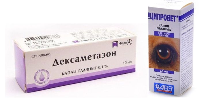
- DexamethasonePlace 2 drops into the corner of the eye 2-3 times daily. Do not use if there are purulent formations in the third eyelid area. A burning sensation may be felt for several minutes after application, but this will subside as the medication is absorbed into the tissue.
- TsiprovetThe drops have an antibacterial effect and are instilled 1 drop four times a day at equal intervals.
Important: folk remedies include oak bark tinctures, chamomile, marigold, St. John's wort, and other herbs with anti-inflammatory properties. Eyes can be treated with cotton pads soaked in the warm solution.
Treatment for third eyelid pathology is divided into conservative and surgical. These two treatment methods are often combined.
- A conservative approach is based on the use of antiseptics (for rinsing the eye), anti-inflammatory agents (to prevent suppuration of damaged tissue), and corticosteroids. Tetracycline ointment, applied to the inflamed areas, has also proven effective.
- Surgical intervention. The procedure is performed under general anesthesia and involves fixing the lacrimal gland to the periosteum of the zygomatic bone. If successful, the integrity of the third eyelid is completely preserved, with no loss of mobility. In most cases, recurrence of eyelid prolapse is 90% unlikely. The dog recovers within 10-15 days.
Important: To avoid scratching and introducing new infections, it is advisable to put a special collar on the animal.If the inflammation is severe before surgery, preparatory therapy may be prescribed, including a course of antibiotics or antimicrobials. Medications should be taken strictly according to the doctor's prescription.
Read also:


Add a comment