
Vasculitis in dogs: symptoms and treatment
Vasculitis is an inflammatory process that affects the walls of blood vessels, leading to disruption of their blood supply. Vasculitis itself is not a diagnosis; it is a symptom complex that can be caused by a number of different factors. Many animals are susceptible to this condition, but it is diagnosed more frequently in dogs than, for example, in cats.
Causes of vasculitis in dogs
There are primary and secondary types of vasculitis. Primary vasculitis is caused by hereditary factors. Breeds predisposed to this condition include German Shepherds, Greyhounds, Dachshunds, Terriers, St. Bernards, and Shar-Pei.
The development of secondary vasculitis can be caused by factors that activate the immune system:
- infectious diseases;
- diabetes mellitus;
- autoimmune disease lupus erythematosus;
- neoplasms.
Vasculitis can be caused by allergic reactions. They develop to medications (itraconazole, prednisolone, dexamtetazone), and the rabies vaccine. The most common food allergens that cause atopic vasculitis in dogs are beef, chicken, lamb, wheat, soy, and corn. Neutrophils produced as a result of the immune system's response to the allergen damage the walls of microvessels, leading to the formation of blood clots, necrosis, and ulcers.
Clinical features of vasculitis
Vasculitis can be cutaneous or systemic. In the former case, the pathology is localized and primarily affects the microvessels of the dermis. In the latter case, vasculitis in dogs affects the eyes, vessels of the kidneys, liver, stomach, intestines, and muscles and joints. The clinical manifestations of vasculitis largely depend on the affected area.
Proliferative thrombovascular vasculitis in dogs develops on the ears, nasal planum, lips, tip of the tail, and, less commonly, on the elbows, footpads, and hocks. Initially, the lesion begins with erythema (redness, rash) that rapidly progresses to form deep ulcers up to 5 cm in diameter that merge into clusters. Photos of dogs with this form of vasculitis show swelling, scaling, papules, erosions, ulcers, hemorrhages, and localized alopecia (hair loss in individual areas).
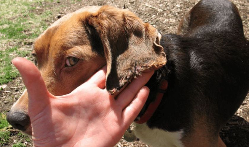
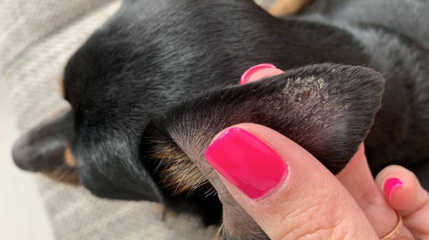
When vasculitis develops in the ears, alopecia of the auricle is first observed, followed by darkening, ulceration, and localized necrosis (the spontaneous dissolution of dead cells). Without proper treatment, the animal may lose its ears. With hereditary cutaneous vasculopathy, enlarged lymph nodes and oral lesions are likely.
Symptoms of vasculitis in dogs with renal glomerular vasculopathy include general depression, increased body temperature, loss of appetite, thirst, polyuria (increased urine production), vomiting, diarrhea, and, in severe cases, acute renal failure, which causes the accumulation of nitrogenous compounds in the blood and often leads to the death of the animal.
Diagnostics
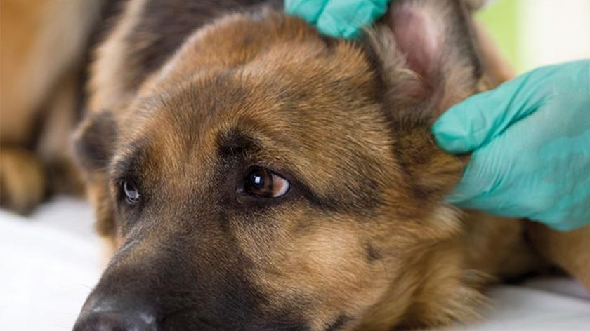
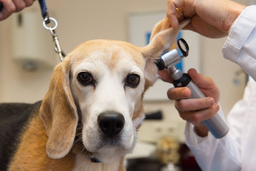
As with any other disease, diagnosis of a vascular disorder such as vasculitis begins with an examination of the sick animal and a study of the anamnesis provided by its owner.
To decide how to treat vasculitis in dogs, it is necessary to differentiate it from other pathologies with similar clinical manifestations: SLE (lupus erythematosus), disseminated intravascular coagulation syndrome, bacterial folliculitis, dermatophytosis (a fungal infection of keratin in the skin), frostbite, and demodicosis (a skin disease caused by mites).
An accurate diagnosis is made based on data obtained from a series of laboratory tests:
- Histological analysis of affected tissue (examination of a tissue fragment under an optical microscope). This method reveals inflammation in the vascular wall, fibrosis (proliferation of connective tissue with scarring), and thrombus formation.
- Direct immunofluorescence analysis of an intact (undamaged) skin sample. This method allows for detailed analysis of biological samples for the presence of specific antigenic determinants and is used to detect deposits of immunoglobulins (antibodies) and immune complexes. The study can be performed manually using a fluorescence microscope or an automated cytometer.
In some cases, a biopsy of a sample of the affected skin is necessary to make an accurate diagnosis.
Treatment
Treatment of vasculitis in dogs is aimed at eliminating the underlying cause. It is performed under the guidance and supervision of a veterinary dermatologist.
In the vast majority of cases, medications are needed to reduce abnormal immune activity and treat autoimmune diseases. Due to their immunosuppressant properties, the following are used in dermatological practice:
- Glucocorticoids Prednisolone, Dexamethasone, Polcortolon, Kenalog, Diprospan, Flosteron, Metypred, Solumedrol, Cyclosporine, Mycophenolate, Aazathioprine, Tacrolimus.
- Cytostatics Doxorubicin, Fluorouracil, Hydroxyurea, Cyclophosphamide, Azathioprine).
- To correct microcirculation and improve blood circulation in microvessels, and reduce platelet aggregation, the following angioprotectors are used: Parmidin, Etamsylate, Calcium Dobesilate, Tribenoside, Troxevasin, Aescusan, Reparil, Esflazid, Pentoxifylline.
In cases of ulceration and bleeding, potent topical immunosuppressants may be necessary. To eliminate secondary infections in dogs with cutaneous vasculitis, ointments, emulsions, and suspensions containing anti-inflammatory steroid components are used.
In mild cases of vasculitis, the prognosis is generally good. If the underlying cause of the vasculitis cannot be eliminated, the dog will require lifelong medication.
Read also:
- Fistula in a dog: how to treat it
- Histiocytoma in Dogs: Causes and Treatment
- Pododermatitis on the paws of dogs: symptoms and treatment


Add a comment