
A lump under a dog's skin: causes and treatment
What should you do if you find a lump or bump under your dog's skin? Of course, don't panic right away, but try to first analyze the possible causes. It could be the result of an injury, a recent vaccination, bee sting And so on. Timely diagnosis and proper treatment will quickly get your pet back on its feet and, at the same time, encourage owners to schedule mandatory veterinary checkups.

General symptoms
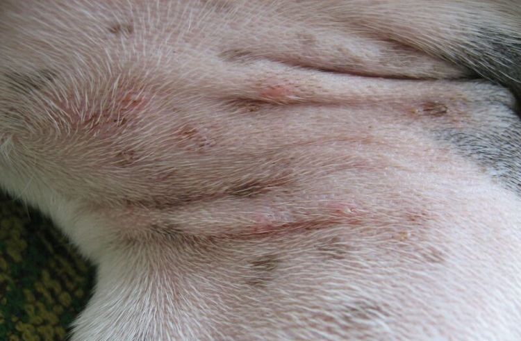
Subcutaneous lumps typically don't bother dogs, even if they're actively growing. Depending on the cause, these spherical bumps can vary in density and diameter, from a few millimeters to several centimeters. Skin irritation and itching are typically caused by insect bites. In other cases, the bump can only be detected by touch. In short-haired breeds, these "defects" are noticeable visually, but in long-haired breeds, they can only be diagnosed by palpation.
If the lump's appearance is suspicious and the lump under the skin does not resolve within 7-10 days, the dog should be examined by a veterinarian. The following symptoms may alert the pet owner:
- The dog reacts painfully to palpation of the lump.
- The lump “grows” before our eyes, increasing in size several times in a short time.
- The subcutaneous tissue opens, and pus or other discharge begins to ooze from the wound.
- The color of the skin around the lump changes.
- The pet becomes apathetic, eats poorly or refuses food altogether, and sleeps restlessly.

Types
Conventionally, all neoplasms can be divided into two groups: benign and malignant. The former include:
- An abscess appears as a lump filled with pus. It can result from a bruise, a fall, a poorly administered injection, or other skin damage. A distinction is made between superficial and deep abscesses. A superficial abscess forms in the upper subcutaneous layer and is easily detected by the swelling and reddening of the tissue. A deep abscess affects internal organs and is not visually detectable.
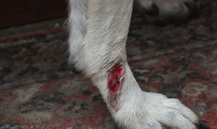
Dogs often experience inflammation of the anal glands, which results in the appearance of lumps around the anus. When the problem is aggravated by infection, the area becomes severely swollen, accompanied by an unpleasant odor.
- Papillomas Warts and warts are common in smooth-haired dogs. The disease is transmitted through contact with a carrier of the virus. The growths can appear singly or in clusters. They hang in clusters on specific areas of the body, have a branched structure, and are dark in color. They are soft and crumbly to the touch.
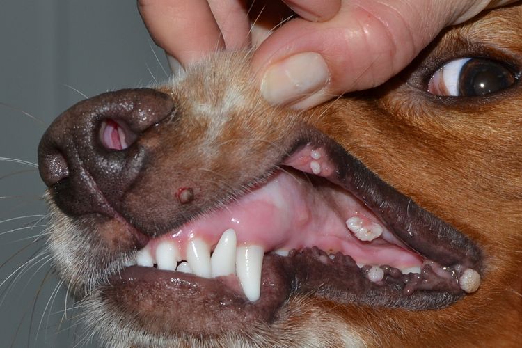
Papillomas typically appear on mucous membranes (mouth, eyes), as well as on the abdomen, groin, and armpits. Despite their unsightly appearance, these hanging "balls" may not cause any discomfort to the animal, but if they begin to change color or bleed, you should see a veterinarian immediately.
- HematomaThey can be caused by ruptured blood vessels and, as a result, hemorrhage into adjacent tissues. The accumulation of excess fluid leads to the formation of lumps, which can resolve spontaneously or require surgical drainage.
Most often, hematomas result from bruises or blows, as well as various medical procedures. The more severe the damage to the vessel, the larger the lump. The dog may react to the problem with a fever, poor appetite, nervousness, or, conversely, apathy. Sometimes, swollen lymph nodes may be observed.
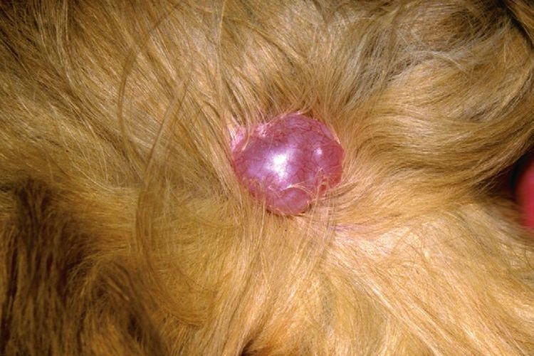
- A cyst. It is always located in the superficial layer of the skin, for example on the back, withers, or muzzle. It is round in shape and usually does not cause pain when pressed. It can range in size from 2-5 cm in diameter and feels loose and soft to the touch. A cyst-like ball can be found under the skin anywhere due to blocked gland ducts. Depending on its location, a cyst may cause no discomfort to the dog or, on the contrary, interfere with movement, chewing, lying on its side, etc.
- PyodermaPuppies up to 4 months of age are susceptible to the disease. Hard bumps appear suddenly and spontaneously. They quickly spread throughout the body, developing into purulent boils. They often spontaneously rupture and become fistula shape. Accompanied by itching and scabies.
The most vulnerable to pyoderma are small breed dogs (Chihuahua, Yorkshire Terrier), as well as boxers, shar pei, and French bulldogs.
Insect bites—spider, wasp, ant, and hornet bites—fall into a separate category. The bite site causes severe swelling, which is painful, itchy, and irritating. Because the dog scratches the affected area with its paws, for example, on the ear, head, scruff, and other areas, the bump may take a long time to heal and develop into an open wound.
The worst-case scenario is the transformation of benign tumors into malignant ones. The rate of progression is impossible to predict; each case is individual. Lumps may remain dormant for years, then begin to change shape, or they may actively grow and immediately become a source of metastases throughout the body.
Older animals are at a higher risk of developing sarcoma, while fibrosarcomas can affect young puppies as young as 6 months. Visually, malignant tumors can only be detected in the late stages.
Diagnostics
When planning a veterinary appointment, it's a good idea to prepare in advance to avoid wasting time and clearly answer the following questions:
- How long has the problem been present?
- Has the appearance of the cone and its size changed, and if so, how, and how quickly it grows.
- How many of these balls were found on the body, and in what order.
- How is the dog behaving? Has its behavior, food preferences, activity level, weight, etc. changed?
- How does the animal react to the lump - does it scratch, lick, not allow you to touch it, or, on the contrary, is indifferent.
- Who the dog has been in contact with recently, at home and outside.
- Was any self-medication carried out: injections, lotions, ointments, etc.
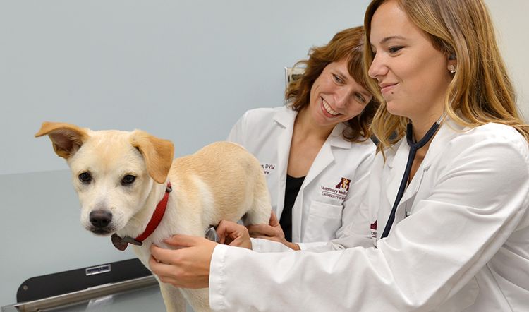
If, after a visual examination, the doctor still has any doubts about the diagnosis, he or she may prescribe additional tests:
- A biopsy is performed by inserting a sterile needle into the lump to obtain a sample. The sample is then sent for testing to determine whether the lump is benign or malignant. This is performed under local anesthesia.
- A smear. This is taken when the lump has broken open and developed into an ulcer. A laboratory slide is placed on the affected area, and the imprint is then examined by a pathologist. The results are available within a few days.
- Computed tomography (CT) scan. This is performed if the subcutaneous tumor is too deep for a biopsy to be possible. CT scans are also used to detect metastases.
- Radiography. An indispensable tool for diagnosing tumors located in the deep layers of the skin.
Read also:
- Lumps on a dog's hind legs: causes and treatment
- Dog Paw Abscess: Cause and Treatment
- My dog's paw hurts after an injection: why and what to do


Add a comment