
Dry Eye Syndrome in Dogs: Causes and Treatment
Dry eye syndrome (keratoconjunctivitis sicca) is a chronic condition common in dogs. It develops due to dysfunction of the lacrimal glands, resulting in insufficient tear production and loss of the protective film on the ocular surface. Left untreated, the condition can lead to serious complications, including vision loss.
This problem has become widespread recently and is therefore actively studied in veterinary ophthalmology.
Reasons
Dry eye syndrome can develop in dogs for a number of reasons. It can be either an independent condition or a consequence of another medical condition.
Here is a list of possible reasons:
- hereditary predisposition;
- poor nutrition (lack of vitamin B12, ascorbic acid);
- age-related changes in the eyes;
- autoimmune diseases;
- herpes infection;
- removal of the third eyelid or Gardner's gland;
- use of drugs with toxic effects (non-steroidal anti-inflammatory drugs and sulfonamides);
- trauma to the lacrimal gland;
- congenital anomalies of the lacrimal gland;
- eye tumors;
- physiological exophthalmos (in pugs, Pekingese and some other breeds);
- "plague of carnivores";
- leishmaniasis.
Symptoms
Keratoconjunctivitis sicca occurs with varying degrees of severity. Diagnosing the condition is difficult, especially in its early stages. Often, owners mistake dry eye syndrome for simple conjunctivitis. Even a specialist can have a difficult time making the correct diagnosis.
Specific signs:
- redness and swelling of the conjunctiva;
- frequent blinking;
- corneal pallor;
- a sharp decrease in lacrimation.
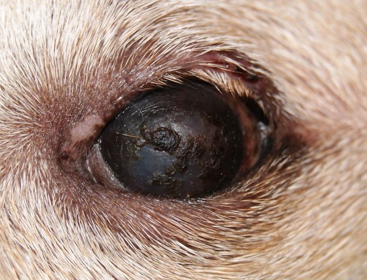
As the condition progresses, characteristic yellowish-greenish discharge with a viscous consistency appears. It is difficult to remove from the eyes. Blepharospasm (involuntary closure of the eyelids) occurs.
In its advanced stages, the disease causes significant discomfort to the dog. In some cases, characteristic ulcers appear, which can vary in size. Vascular disease develops, and later pigmentary keratitisBrown pigment is deposited in the layers of the cornea, and the animal gradually loses its vision.
Diagnostics
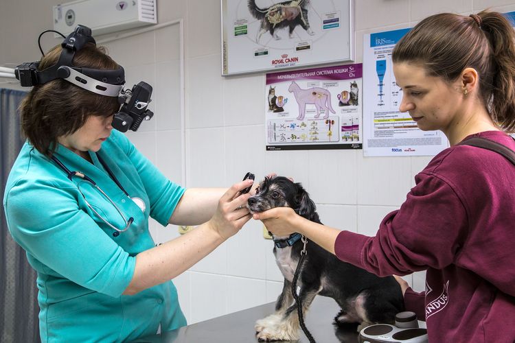
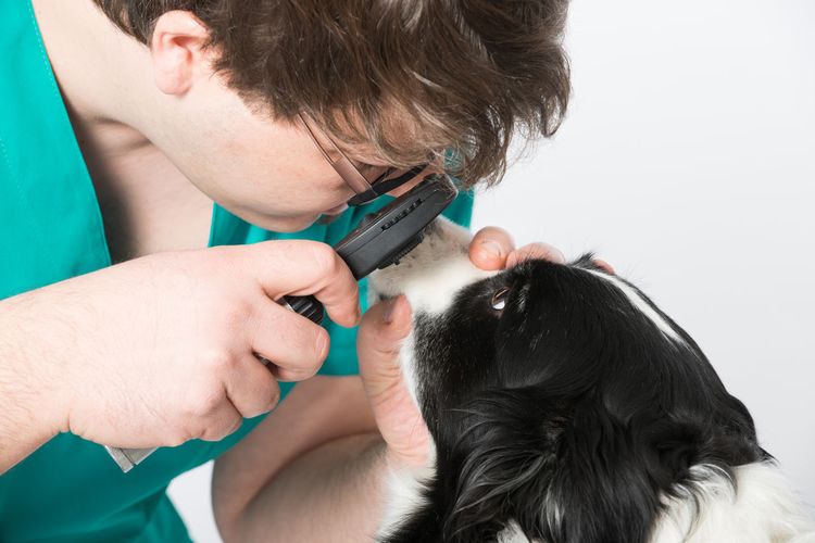
First, the specialist carefully examines the animal, assessing the condition and position of the eyelids, and the nature of any ocular discharge. A medical history of previous injuries, surgeries, and illnesses is also collected.
If dry conjunctivitis is suspected, the veterinarian will prescribe special tests to confirm or refute the diagnosis.
To determine the number of dead corneal cells, specific dyes (such as a 1% fluorescein solution) are used. The dye highlights defective areas, even if the epithelial cells are only slightly altered.
Other diagnostic methods:
- The Norn functional test provides information on the stability of the tear film.
- The Schirmer test is used to measure tear production. The amount of tears secreted per minute is measured using filter paper strips. They are applied to the lower eyelid, and the wetted length is measured after removal.
- Blood test for hormones.
- Biochemical blood test.
- Bacteriological culture (in complicated cases).
Treatment
If your dog has dry eye syndrome, it's crucial to consult a veterinarian promptly and begin treatment. Treatment can be conservative or surgical. The extent of therapy depends on the severity of the condition. A comprehensive approach is always used.
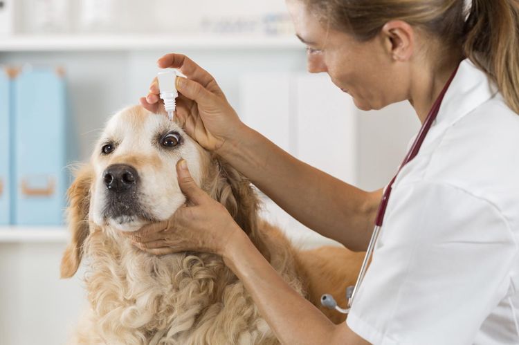
Standard therapy involves the use of an artificial tear stimulator. It is selected individually for each case and is used until tear production is fully restored. There are numerous medications available on the market that moisturize the ocular surface and form a stable film. Pigmentation gradually fades, the cornea becomes transparent, and vision is restored.
List of drugs:
- Oftagel;
- Vidisik;
- Lacrisin;
- A natural tear.
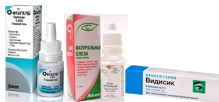
The lower the viscosity of the drug, the more often it needs to be used.
It is also necessary to eliminate the clinical symptoms of the disease. Signs of purulent conjunctivitis and eyelid spasms resolve within 7-14 days. The next step involves prescribing antibacterial medications to control secondary microflora.
Sometimes it's helpful to use homeopathic remedies. They stimulate tear production. These are typically eye drops. The recommended dosage is 5-10 drops twice daily.
Other adjuvants include keratoprotectors (corneal protectors):
- Actovegin;
- Solcoseryl;
- Korneregel.
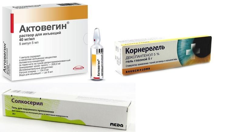
They improve metabolic processes, accelerate tissue regeneration, and eliminate discomfort. Significant improvements are noted after just 3-5 days of use.
If necessary, local antihistamines are used:
- Allergodil;
- Spersallerg.
Even after full recovery, you should continue to have regular veterinary checkups (a couple of times a month). During these visits, the veterinarian will perform monitoring measurements to determine whether tear production is normal.
In some cases, surgical intervention is necessary. Possible options:
- Partial tarsorrhaphy. This is a simple procedure aimed at reducing the palpebral fissure.
- Repositioning the parotid duct. This will ensure that the eyes are moisturized by salivary fluid.
- Removal of the superficial layer of the cornea. This is a complex and traumatic procedure. It is used only when tear production is normal.
Forecast
If keratoconjunctivitis sicca is caused by an immune-mediated disorder, lifelong therapy will be required. In other cases, treatment is continued until the lacrimal glands fully recover. In any case, strict adherence to the doctor's recommendations is the key to success.
It's important to always closely monitor your pet's eye condition. Early diagnosis will help eliminate the problem as quickly as possible.
Read also:
- My dog has red eyes: why and what to do
- Inflammation of the third eyelid in dogs: symptoms and treatment
- Treatment for a Dog's Eye Spot


Add a comment