
Umbilical Hernia in Dogs: Causes and Treatment
An umbilical hernia can occur in dogs of any breed. Owners may notice this characteristic bulge on a puppy's abdomen both in the first days of life and later in life. In this article, we'll examine the main causes of this problem in detail, explain how to treat it, and what treatment a veterinarian may recommend.
Content
Hernia in a dog
A hernia is a prolapse of soft tissue or organs located in the abdominal cavity through a pathological opening that forms in anatomical structures under the influence of various factors.
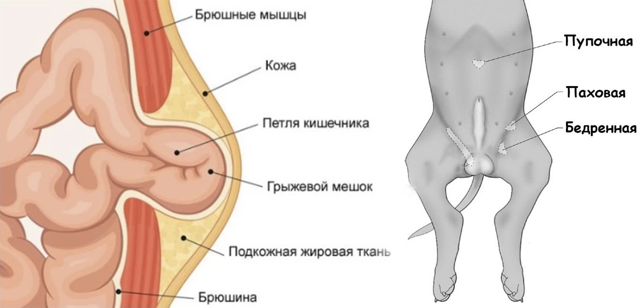
Depending on the area of the opening, the hernia may be internal (the protrusion occurs into the cavity and is not visualized) and external (the hernia is visible on the dog's belly).
Diagnosing internal hernias is complicated by the lack of the primary symptom—the characteristic protrusion. A doctor may discover a protrusion of the esophagus, sigmoid colon, cecum, or rectum (or even strangulation) only during a comprehensive examination.
Among the external hernias visualized on the animal’s abdomen, the most common are:
- umbilical;
- inguinal;
- femoral;
- perineal.
Moreover, an umbilical hernia in dogs is often diagnosed in early puppyhood, while an inguinal or perineal hernia can occur in dogs at any age.
There is also intervertebral hernia, which has a slightly different nature than the previously described types. We previously discussed how spinal disc herniation in dogs is diagnosed and treated in more detail in a related article.
This article will focus specifically on umbilical hernias, which many breeders mistakenly consider a harmless condition that requires no treatment, while veterinarians consider them a serious condition that should be treated as quickly as possible.
Symptoms of an umbilical hernia
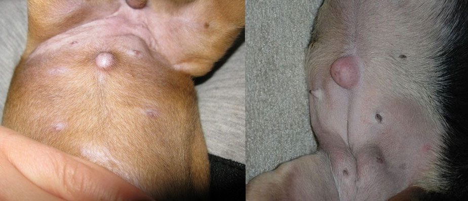
An umbilical hernia in a puppy is difficult to confuse with other types of pathology, as it looks like a characteristic painless protrusion in the navel area.
Depending on the size of the opening and the contents that fall out (omentum, intestinal loop, fragment of the bladder or uterus), the following types of hernias are distinguished:
- False (a small, bulging area, soft to the touch, easily repositioned, no organ prolapse). If left untreated, the sac may persist and fill with fatty tissue.
- True (The organ prolapses through the opening.) True hernias, in turn, can be reducible, non-reducible, or strangulated.
An umbilical hernia in a puppy looks like this.
When discussing whether an umbilical hernia is dangerous for a puppy, many breeders point to false hernias, which are quite common in practice. Veterinarians, however, always evaluate potential complications, such as necrosis of the strangulated area, as well as inflammation, which, without proper treatment, can lead to sepsis and death.
At home, it is very difficult to determine the type of hernia in a puppy and its danger based only on external symptoms.
When assessing whether your pet's condition is dangerous, it is important to remember that symptoms that require emergency care include:
- a protrusion that does not return to normal with gentle pressure;
- compaction of the hernia (indicates that there is strangulation);
- an increase in the size of the protrusion (may indicate swelling of the pinched area);
- redness and inflammation in the area of the protrusion;
- deterioration of the dog's general condition (depression, refusal to eat, vomiting, constipation).
Causes of the pathology
An umbilical hernia is often congenital. Prolapse of the omentum or intestinal loop occurs through an overly large opening in the unclosed umbilical cord.
Normally, the opening should close postnatally, when the blood vessels that nourish the fetus through the umbilical cord cease functioning. However, if this doesn't happen, the puppy develops an umbilical hernia.
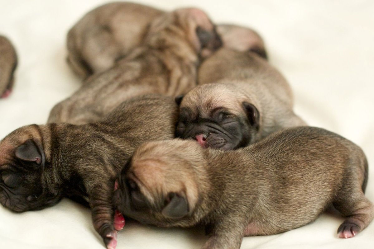
It's believed that small-breed puppies are more prone to developing umbilical hernias in the first weeks of life. Veterinarians often diagnose this condition in Chihuahuas, Shih Tzus, Yorkies, as well as young Pugs and French Bulldogs.
But it's important to understand that the problem can also occur in adult animals of any breed (Dachshunds, Pit Bulls, German Shepherds, etc.). The main causes of umbilical hernias in puppies over 1 month old or adult dogs are:
- abdominal trauma;
- abdominal muscle strain;
- excess intra-abdominal pressure.
Diagnosis and treatment methods for umbilical hernia
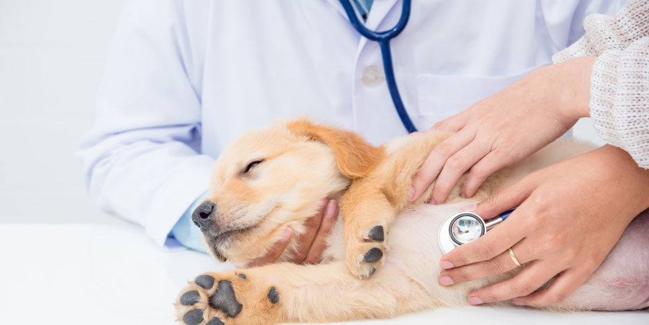
The doctor determines how to treat a hernia based on the results of a comprehensive examination, which may include not only a visual examination, but also a series of tests, an abdominal ultrasound, an X-ray (with contrast), and an assessment of the heart's condition (since general anesthesia is required for surgery).
The treatment method recommended by your veterinarian will depend on many factors:
- A type of hernia.
- Age of the animal.
- General condition of the dog.
Method No. 1 - gluing
This technique is effective in newborn puppies up to 1 month old and is used for false umbilical hernias or uncomplicated reducible true hernias with a small ring diameter.
Important! Do not attempt to reduce a hernia at home. This procedure should be performed by a veterinarian after examining the animal.
Method No. 2 – surgical treatment
Umbilical ring suturing surgery is the most effective method for treating umbilical hernia.
Many veterinarians believe that it's best to perform surgery immediately rather than trying to patch a congenital hernia. Once the underlying cause is surgically removed, the puppy will be able to develop fully and lead an active lifestyle. Furthermore, the sooner the hernia is removed, the fewer health consequences veterinarians predict for the animal.
- Timely removal of a false or uncomplicated hernia guarantees a 100% recovery without adverse health consequences for the dog, with one exception. Dogs with a history of congenital umbilical hernias should be excluded from breeding!
- If a choke occurs, there's a high risk of necrosis developing. In this case, excision of the dead tissue is necessary, which could have long-term consequences for the animal's life.
- The most dangerous scenario is the development of peritonitis. In complicated cases, the prognosis is guarded, and even with prompt surgery, a fatal outcome cannot be ruled out.
The optimal option is when umbilical, inguinal, or perineal hernias in dogs are surgically performed on a planned basis (before dangerous symptoms develop). In this case, the surgery itself is quick and less traumatic, and the recovery period is shorter.
Simultaneous castration (sterilization) and suturing of uncomplicated umbilical hernia is permitted.
Postoperative period
For planned uncomplicated hernia closure, the animal's postoperative rehabilitation takes place at home. You can take your operated dog home after recovery from anesthesia.
Caring for the seam at home includes:
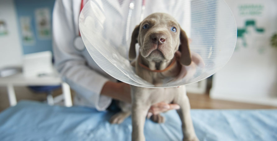
- wearing a collar or blanket to prevent the seam from being licked;
- treatment of the suture with antiseptic solutions (as recommended by a veterinarian);
- application of antibacterial ointment (as recommended by a veterinarian).
If emergency surgery was performed following complications (strangulation, tissue necrosis, peritonitis, sepsis), the animal may require around-the-clock medical care for several days after surgery to stabilize its condition. During this period, the dog will remain in the clinic. Treatment will include intravenous antibiotics, IV fluids with supportive medications (depending on the severity of the condition), treatment for dehydration, and constant monitoring of vital signs to ensure prompt response if the condition worsens.
Veterinarian's advice
Read also:
- Gingivitis in Dogs: Causes and Treatment
- Hot Ears in Dogs: Why and What to Do
- White and Pale Gums in Dogs: Causes and Treatment


Add a comment