
Corneal abrasion in dogs: treatment
The cornea is the outermost layer of the eye, composed of fibrinous fibers. In dogs, this layer is approximately 0.5 mm thick and ranges from 12 to 18 mm in diameter. Smooth, transparent, and avascular, the convex cornea occupies almost the entire visible portion of the eye, providing 70% of light refraction and providing protection. Corneal injuries are a common problem in dogs, as these animals are active, inquisitive, and not always cautious. Dog owners should know how to provide first aid to their pet and how to treat corneal injuries.

Content
Causes of corneal damage
Corneal injuries are most common in hunting dogs, due to their professional activities: a dog can injure an eye while wading through thorny bushes or during a fight with another dog of the same breed. Eye injuries can also be caused by:
- wire and other sharp objects;
- chemicals;
- cat's claw (this is the most dangerous type of injury, a cat's claw can cause a deep wound, and there is also a large amount of pathogenic microflora under it);
- Ectopia (eyelashes that grow toward the cornea) or entropion (inward turning of the eyelid). In these conditions, the eyelashes constantly irritate the eyeball.
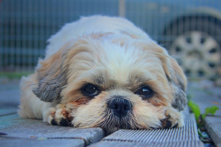
Dog breeds with bulging eyes and flat muzzles (brachycephalic) are at risk for corneal injuries, as are breeds with a genetic predisposition to entropion or ectopia. These include St. Bernards, Rottweilers, Labradors, Boxers, English Bulldogs, Chow Chows, and, among small breeds, the Pekingese and Shih Tzu.
Symptoms
The cornea contains many nerve endings, so when it's damaged, dogs experience discomfort and pain. They become restless, often rubbing the injured eye with their paw or against objects. Symptoms that indicate corneal injury also include:
- profuse lacrimation;
- redness of the conjunctiva of the eye;
- clouding of the cornea in the damaged area;
- spasm of the eyelids (blepharospasm);
- photophobia (the animal turns away from the light source, squints, closes its eyes).
Caution! Self-treatment, including removing a foreign object lodged in the cornea, is strictly prohibited. Such manipulations can cause a rupture of the cornea and damage to the integrity of the eyeball. However, first aid can and should be administered to the injured pet, after which the dog should be transported to a veterinary clinic as quickly as possible.
First aid
First of all, you should prevent your dog from scratching the injured eye. To do this, place a protective collar on your dog. These devices are sold at veterinary pharmacies and are truncated cones made of clear plastic, the narrow end of which attaches to the collar. At home, you can make a protective collar out of cardboard, very thick fabric, or a plastic kitchen towel. The diameter of the bottom of the collar should match the size of the collar when fastened, and the width should be sufficient to cover the dog's entire neck and head, down to the tip of the nose. The top and bottom edges of the collar should be wrapped with medical tape.
If it takes a long time to get to the vet, you can ease your pet's discomfort by placing 2% novocaine or inocaine drops in the eye. These are local anesthetics that take effect within 0.5-1 minute and last for up to half an hour. Other eye drops or ointments, as well as folk remedies, cannot be used without a veterinarian’s prescription.
Diagnostics
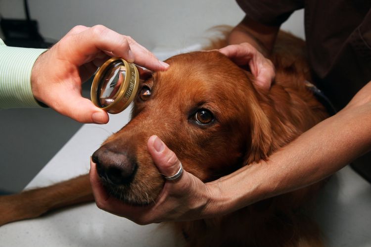
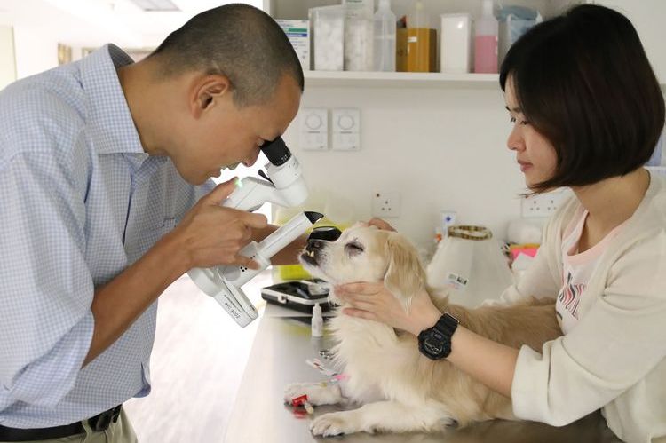
To determine the cause and assess the extent of corneal damage, a veterinary ophthalmologist performs a physical examination of the dog. They then examine the cornea with a slit lamp and magnifying glass (foreign bodies are often so small that they cannot be detected without such a device).
The animal also undergoes a water-based dye test (fluorescein is commonly used). The damaged area is highly hydrophilic and therefore stains during the test. If necessary, the dog may undergo an ocular ultrasound. Due to the somewhat painful nature of the diagnostic procedures, local anesthesia or sedation are used to reduce pain.
Treatment
Treatment for a damaged cornea in a dog depends on the severity of the injury. For superficial corneal damage, medications that reduce inflammation and eliminate pathogenic microflora are sufficient.
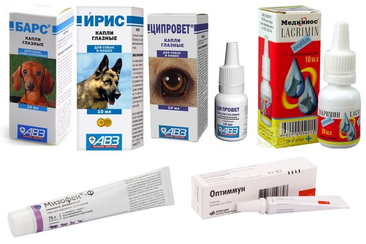
In veterinary medicine, eye drops are used for this purpose:
- «Leopard» (active ingredients: chloramphenicol and furacilin);
- "Iris" (active ingredient gentamicin sulfate);
- "Tsiprovet" (active ingredient ciprofloxacin);
- "Lacrim" (active ingredients: chloramphenicol and sodium sulfosalicylate);
Your veterinarian may also prescribe eye ointments for your dog, which should be applied under the animal's lower eyelid. Ointment-based medications have a good anti-inflammatory and disinfectant effect:
- "Mizofen" (active ingredients chloramphenicol and levamisole hydrochloride);
- «Optimum" (active ingredient cyclosporine);
- Oxytetracycline ophthalmic ointment.
In case of extensive or deep damage to the cornea, it is necessary to resort to surgical treatment: optical reconstructive surgery or transplantation of a donor cornea.
Important! To prevent your dog from aggravating the injury or introducing an infection by scratching the affected eye, wear a protective collar for the entire treatment period. Its design allows the animal to see normally and does not interfere with eating.
Preventing eye injuries in dogs
To minimize the risk of corneal damage, it's recommended to walk your dog on a leash. This will prevent it from getting into street fights or injuring its eye while running through bushes. When traveling by car, don't allow your dog to stick its head out the window.
If you have both dogs and cats in your home, ensure they coexist peacefully. Keep all household chemicals out of reach of your pets.
Read also:
- Third eyelid prolapse in dogs
- Dry Eye Syndrome in Dogs: Causes and Treatment
- Inflammation of the third eyelid in dogs: symptoms and treatment


Add a comment