
Mastocytoma in Dogs: Symptoms and Treatment
Mastocytoma in dogs is an abnormal proliferation of mast cells (mastocytes). These cells are located in connective tissues and are involved in immune processes and the transport of immunoglobulins. Small, seemingly harmless tumors can quickly transform from a stable state into a malignant tumor, causing metastases to other organs. Early detection and proper treatment can significantly prolong the pet's life.
Content
Reasons for development
Veterinarians still cannot determine the exact factors that trigger the development of mastocytoma. Some hypothesize that the condition develops due to exposure to viruses and adverse environmental factors. Furthermore, most veterinarians agree that its development is preceded by a mutation in the proto-oncogene gene, which is involved in hematopoiesis and mast cell proliferation. These tumors can subsequently develop into tumors, which is why they are also called mast cell tumors.
Risk group
Mastocytoma is one of the most common types tumors in dogs (accounting for up to 20% of all skin neoplasm diagnoses), it can develop regardless of age, gender, or breed. The disease is most common in the following breeds:
- bulldogs;
- boxers;
- Staffordshire Terriers;
- pit bull terriers;
- bullmastiffs;
- beagles;
- Shar Pei;
- dachshunds;
- pugs;
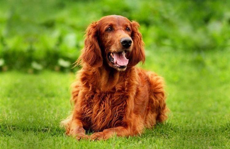
- golden retrievers;
- English Setters.
In addition, about 85% of cases of pathology diagnosis occur in dogs older than 7-8 years.
Symptoms of the disease
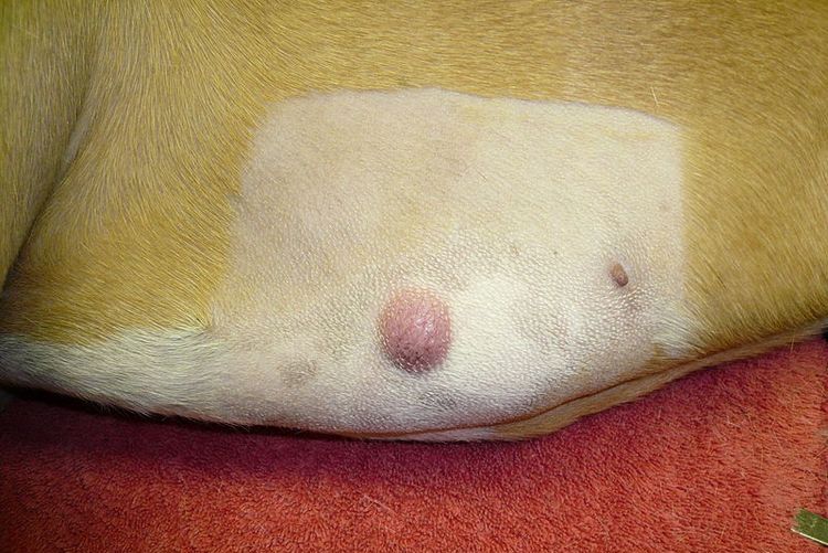
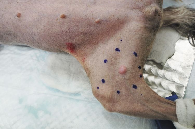
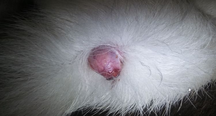
One of the first signs of mastocytoma is a mole-like growth on the skin, papilloma A wart or tumor, where hair loss may occur. It most often appears on the abdomen or extremities, and less commonly on the neck, head, or genitals. In the early stages, the tumor is often characterized by a slow growth rate (if isolated) and often causes no discomfort to the animal. It may have a vague or defined outline, be soft or hard, and be pink to dark cherry in color, or be colorless. The appearance of the following symptoms in a dog should prompt immediate veterinary attention:
- Severe itching, forcing the dog to scratch and chew the affected area.
- Redness, swelling, inflammation at the site of the tumor.
- Erosions and ulcers of the formation area.
- Formation of papules and pustules ranging in size from a few millimeters to 4-5 cm.
- Associated health problems (vomiting, traces of blood in stool) and diseases (duodenal ulcer, blood clotting disorder).
Important! One of the dangerous symptoms of mastocytoma is Darier syndrome. If red nodules form when rubbing the tumor and surrounding skin, this indicates an aggressive stage of development.
Due to the variety of symptoms, veterinarians often refer to mastocytomas as "mimicking tumors," as they can disguise themselves in the early stages as a standard allergic reaction. These tumors can be malignant or benign, and tumors of various etiologies can develop nearby.
The diagnostic process
A mastocytoma should only be diagnosed by a veterinarian after an initial examination of the dog, including necessary tests, X-rays, ultrasound, and histological and cytological examination of the tumor site. Based on these results, the type of pathology is determined:
- Type 1 (approximately 70% of cases) are small, benign growths that appear on the skin or subcutaneous tissue. They are not prone to metastasis and are easy to remove.
- Type 2 – often localized in the subcutaneous tissue and can degenerate into malignant tumors. Even after removal, they can continue to develop with unpredictable consequences.
- Type 3 – develops in the lower layers of subcutaneous tissue and, in the absence of immediate intervention, has an unfavorable prognosis for the animal.
Another goal of diagnosis is to determine the stage of development of the pathology, depending on which treatment will be selected:
- 0 – a single formation in the skin that does not affect the lymph node.
- I – one larger tumor that does not involve the lymph node.
- II – a single tumor with small metastases in the lymph node.
- III – several deep neoplasms, often metastasizing to the lymph nodes.
- IV – single or multiple tumors that metastasize not only to the lymph nodes, but also to the dermal layer.
Treatment methods
Treatment for mastocytoma in dogs depends on the overall clinical picture and the individual characteristics of the animal. The most appropriate method is selected based on these factors:
- Surgical removal is particularly effective for type 1 or 2 tumors, but is contraindicated for multiple or poorly differentiated mastocytomas. Before surgery, a diagnostic resection is performed—pathological tissue is collected to determine the tumor's boundaries. During the surgery, some healthy tissue is removed to minimize the risk of recurrence. Subsequently, the animal requires regular veterinary examinations (every 2.5-3 months).
- Chemotherapy can be used after surgery or instead of it (if surgery is contraindicated). This involves selecting medications that help prevent or delay tumor growth, and sometimes even reduce its size. The most commonly used drug is prednisolone.
Important! Successful treatment of mastocytoma can only be achieved with timely detection, determination of the type, and determination of the stage of development. Therefore, if you notice any suspicious growths on your pet, it is important to promptly consult a veterinarian.
Further forecast
Mast cell tumors are characterized by unpredictable behavior, making it difficult to predict the animal's future life. However, certain conclusions about the chances of recovery can be drawn based on the following factors:
- The degree of differentiation (similarity to normal tissues and cells). A higher degree minimizes the risk of metastasis, meaning the prognosis is favorable. A moderately differentiated tumor can predict a life expectancy of 1-3 years, while poorly differentiated (aggressive) tumors can last up to 12 months.
- Characteristics (size, growth rate, location). Numerous or large, rapidly growing, fuzzy lesions are generally associated with a poor prognosis. In terms of location, lesions on the extremities are considered the most favorable, while lesions on internal organs are considered the least favorable.

Reviews
Natalia, the Labrador's owner:
"Gretta's whole thing started at age 7 with a harmless lump under her jaw. During surgery, the tumor was removed along with a lymph node. She then underwent a course of prednisone, which also went without any negative consequences. She's almost 10 years old, and so far, everything is fine, with no signs of illness. But now, every pimple or mole is immediately taken to the vet."
Sergey, beagle owner:
"The dog developed a large tumor in his lower abdomen. A biopsy revealed a stage 2 mast cell tumor. Due to its large size, the veterinarian did not recommend surgery, as it would require removal of a large amount of surrounding tissue. Treatment offered was a dexamethasone blockade of the tumor. The doctors are not giving a prognosis because they cannot predict its further development, so for now we are continuing treatment and hoping for the best."
Read also:
- A lump under a dog's skin: causes and treatment
- Hypothyroidism in Dogs: Symptoms and Treatment
- Intervertebral disc herniation in dogs


Add a comment