
Keratoconjunctivitis in dogs: symptoms and treatment
Keratoconjunctivitis, or dry eye syndrome, is a common complaint among dog owners. Unfortunately, identifying the symptoms in the early stages can be challenging due to the vague clinical picture, which complicates diagnosis and subsequent treatment.
For normal visual function, the cornea must be kept moist. Lacrimal fluid acts as a protective barrier, washing away foreign bodies and preventing pathogens from entering the eye. It contains substances that nourish the cornea and control the microflora of the fundus. When tear production is disrupted, dry eyes occur, leading to discomfort such as burning, frequent blinking, clumping eyelashes, and other side effects. Failure to seek veterinary attention promptly can lead to serious complications, including vision loss.
Content
Causes of occurrence
Based on medical experience, dry eye syndrome can be both an independent condition and a consequence of another medical condition. The most common causes of dry eye syndrome are considered to be the following:
- Decreased immune system defenses
- Disorders of the nervous system, including those caused by trauma and other mechanical damage to the eye “from the outside”.
- As a complication after general anesthesia, the use of atropine
- Surgical removal of the third eyelid.
- Congenital anomalies. One possibility is the absence of the lacrimal gland altogether or its underdevelopment.
- Chemical and thermal burns of the eyes
- Systemic diseases (distemper, diabetes mellitus, AIT)
- Taking medications that affect the amount of tear fluid produced. These include certain nonsteroidal anti-inflammatory drugs (NSAIDs) and sulfonamides.
- Chronic inflammatory process of the ciliary margin of the eye.
- Herpes
- Age-related changes that lead to a decrease in the function of tear fluid production.
- Poor nutrition, vitamin deficiency.
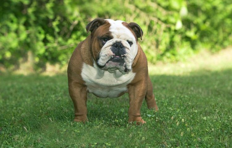
Important: congenital pathologies are most often found in Yorkshire Terriers and Pugs, as well as Poodles, Shih Tzus, and English Bulldogs.
General symptoms
Recognizing the signs of incipient keratoconjunctivitis can be difficult due to the lack of specific clinical signs. Typically, dogs will exhibit:
- increased lacrimation,
- conjunctival edema,
- redness of the conjunctiva of varying intensity,
- small and intermittent purulent discharge from the eyes
The listed symptoms are often associated with conjunctivitis or the result of a foreign body getting into the eye.
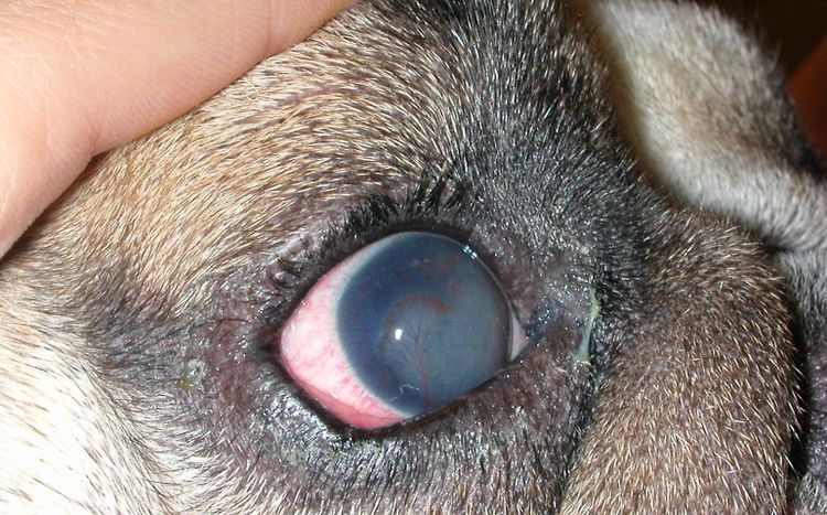
The moderate stage is characterized by obvious signs of decreased tear production and corneal clouding. The following symptoms are also present:
- Profuse purulent discharge from the eyes in the form of viscous mucous threads. The dog has difficulty opening its eyelids, especially after sleep.
- The conjunctiva sticks to the cornea due to the large amount of mucus produced.
- Presence of traces of xerosis (erosion) of the cornea.
- Development pigmentary keratitis of varying degrees of severity.
In advanced cases, obvious degenerative changes in the conjunctiva and cornea are observed, complicated by a persistent decline in lacrimation. Also present are:
- Blepharospasm.
- Purulent discharge from the eyes.
- Damage to the eyelids and subsequently to the skin around the eyes.
- Gluing eyelashes.
- Swelling and inflammation of the conjunctiva
- Changes in the structure of the corneal relief, the appearance of ulcers and perforations.
- Vascular keratitis.
In the final stages of the disease, the animal may permanently lose vision due to complete deformation of the cornea. It becomes not just opaque but covered with a thick, purulent crust.
Diagnostics
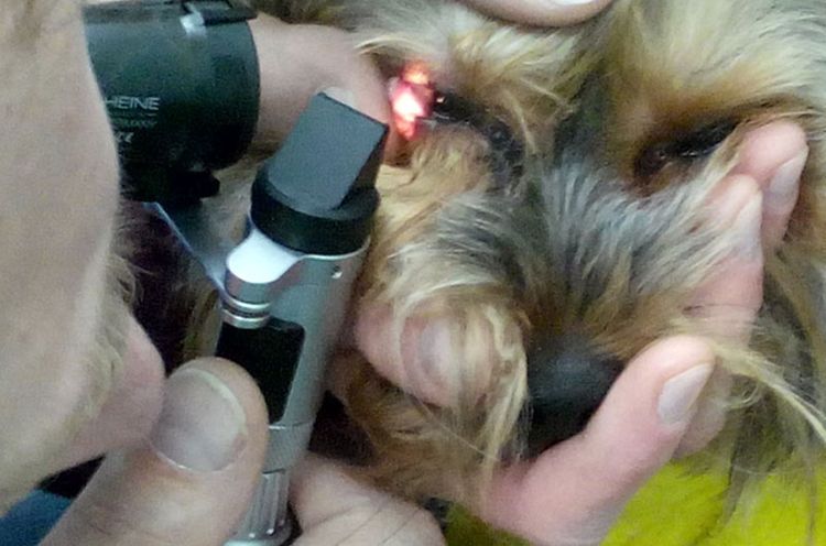
Since it is quite difficult to detect keratoconjunctivitis in the early stages, several tests can be used for diagnosis.
Nornu's Test
Its purpose is to determine the level of tear film stability. To do this, one drop of sodium fluorescein (0.2%) is injected into the lower conjunctival sac and the time between the last blink and the appearance of a black spot (break) on the surface of the tear film is measured.
- less than 5 sec. - critical level;
- 5-10 sec. - below normal;
- more than 10 seconds is normal.
Schirmer's test
This allows one to determine the total volume of tear production. Specially marked strips of filter paper are used for the experiment. The strip is placed at a specific angle in the outer corner of the eye on the edge of the lower eyelid, after which the dog's eye is closed for one minute. After this time, the strip is removed and the length of the area soaked with tear fluid is analyzed.
- less than 5 mm - the maximum level of dry eye;
- below 10 mm - low level of dry eye;
- 11-14 mm - borderline level of dry eye;
- more than 15 mm is normal.

Additionally, blood tests for biochemistry and general examination may be performed. These are relevant if a systemic disease is suspected.
Treatment
Treatment methods for keratoconjunctivitis sicca are divided into therapeutic and surgical. Sometimes, they are combined to achieve maximum effectiveness.
Drug therapy methods include:
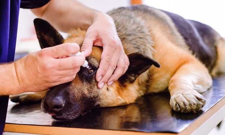
- Artificial tears are used to replace the deficient tear fluid. They are available in liquid or gel form. Depending on the component of the tear film being replaced, the viscosity and chemical composition of the drops will vary. These products perform a common function: moisturizing the surface of the eye, forming a stable film on the cornea. Based on their viscosity, artificial tears are classified into three groups: low (Natural Tears, Hemodez), medium (Lacrisin), and high (Oftagel, Vidisik).
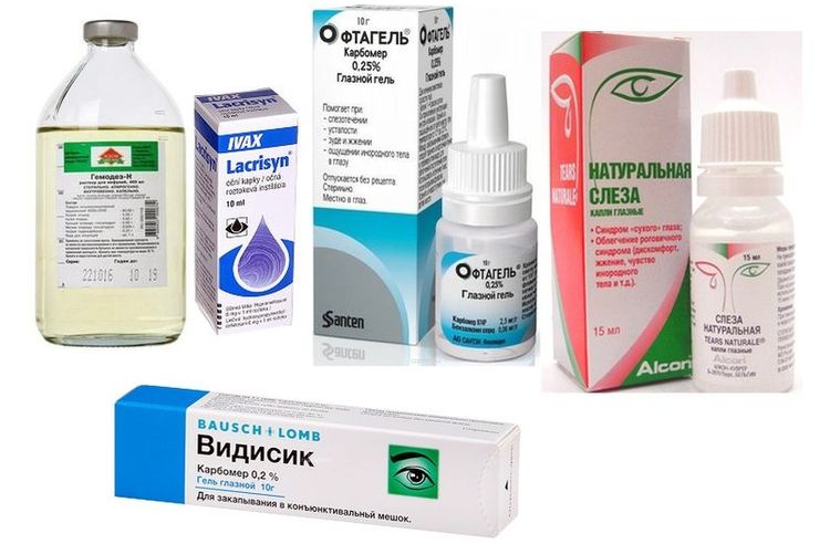
The frequency of instillation depends on the viscosity of the medication. The more liquid the drops, the more frequently they need to be instilled. With low viscosity, the number of applications can reach 5-8 times a day; with high viscosity, only 2-4 times a day.
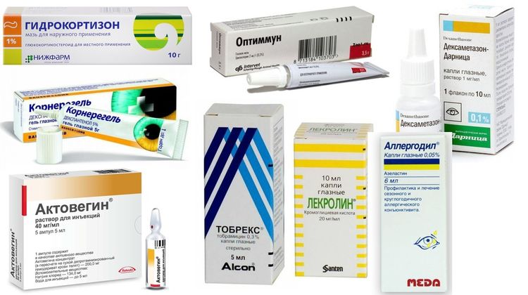
- Increased tear production is regulated with the help of special ointments - medicinal films. Most often, this Optimum (Optemmun) and Cyclosporine-A. Most animals experience a positive reaction, which leads to significant tear production.
- Anti-inflammatory medications: Hydrocortisone ointment, Dexamethasone drops, Prenacid ointment. These medications are not prescribed if there is damage to the corneal epithelium.
- Antibiotics. These are advisable when secondary infections are detected, as well as when there is an imbalance in the secondary microflora. Broad-spectrum antibiotics such as Ciprofloxacin and Tobramycin have proven effective in practice.
- Corneal protectors. They restore tissue metabolism and accelerate regeneration processes. Actovegin and Cornegel are typically prescribed.
- Antihistamines. They are included in the treatment regimen for allergic reactions that trigger dry eye syndrome. These include: Lecrolyn, Spersallerg, Cromoghexal, and Allergodil.
Surgical treatment involves transferring one of the parotid gland ducts to the eye. The procedure is complex and is therefore only performed when drug therapy has proven ineffective.
The function of the parotid gland is to secrete saliva, which then flows through the duct into the oral cavity. Since saliva is almost identical in biochemical composition to tears, it can easily act as a substitute. The duct from the parotid area is relocated to the periorbital area so that saliva flows directly to the eye.
The mineral deposits that build up on the cornea over time are removed using special eye drops. The surgery has a side effect that is harmless but may cause some discomfort to the dog. During feeding, saliva production increases not only in the mouth but also in the eye, so the dog will involuntarily "cry" until it finishes eating.
Keratoconjunctivitis in dogs: video
Read also:
- Inflammation of the third eyelid in dogs: symptoms and treatment
- Treatment for a Dog's Eye Spot
- Why might a dog have red eyes?


Add a comment