
Histiocytoma in Dogs: Causes and Treatment
Histiocytoma is a common skin condition in dogs. It's a benign tumor of the vascular connective tissue, in which Langerhans cells play a key role. Owners often confuse this condition with cancer, giving up on their pet's health. However, by understanding the causes and basic treatment principles, you can help your dog at the early stages of the disease. The key is to catch it early!
Reasons
Histiocytoma in dogs is the result of a mutation in the Langerhans cell genome. Histiocytes, connective tissue cells that are always dormant and then proliferate when inflammation develops, forming a barrier around pathogenic microbes that blocks their further activity, are a normal part of any living organism. Histiocytes act as a marker for inflammation and often even indicate the onset of cancer.
As a result of genetic disorders, exposure to carcinogens, radiation, and a generally weakened immune system, histiocytes begin to mutate and divide uncontrollably. Cutaneous histiocytoma is a disease with the best prognosis of all those associated with histiocyte dysfunction. A single lesion is not alarming and requires competent treatment. Multiple histiocytomas, however, trigger the development of cutaneous Langerhans cell histiocytosis, which can subsequently cause lymphatic damage.
Systemic histiocytosis is diagnosed when generalized lesions of the skin, lymphatic system, and mucous membranes occur. The disease is proliferative in nature, affecting both the outer layers of the dermis and internal organs. It most commonly occurs on the face, eyelids, nose, extremities, and scrotum. The prognosis for this disease ranges from guarded to poor. Systemic histiocytosis is characterized by alternating regression and rapid progression, making it difficult to treat.
Histiocytes that differentiate into interstitial dendritic cells (IDCs) when mutated cause cutaneous histiocytosis of a different etiology, which affects the deep layers of the dermis and subcutaneous tissue.
IDC cells can develop histiocytic sarcoma – a malignant neoplasm that is difficult to treat and has a very unfavorable prognosis.
Cutaneous histiocytoma can occur in any animal, but certain breeds are more likely to develop histiocyte mutations. These include bull terriers, dachshunds, and greyhounds. boxers, Scottish and Boston terriers, Great Danes, smooth-coated retrievers, and cocker spaniels. There is also an age limit: animals under 3 years of age are susceptible to the disease.
Symptoms of the disease
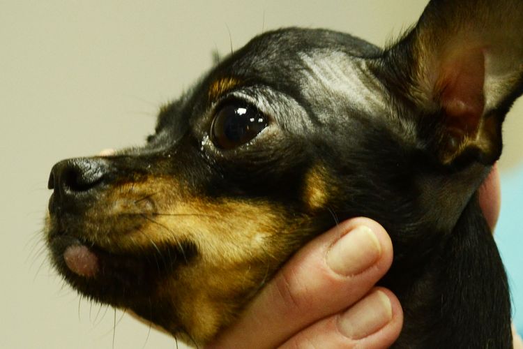
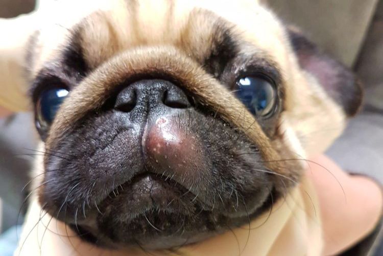
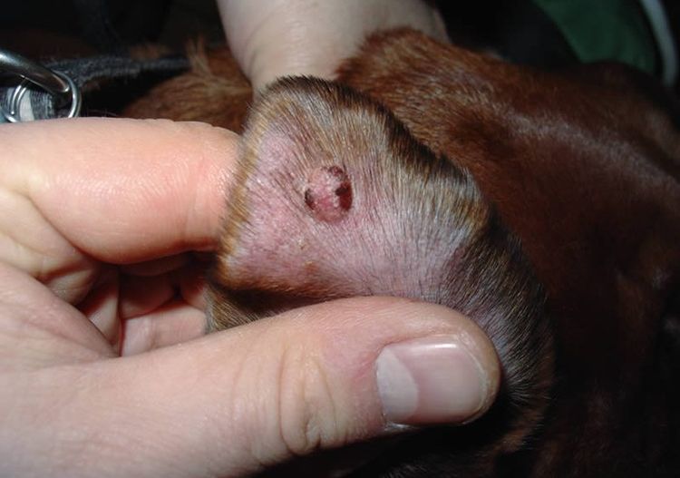
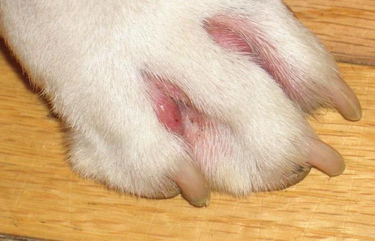
The first sign of a histiocytoma is the appearance of a red spot. Initially, it is flat, but over time, a swelling develops in its place. The growth is firm to the touch, like a ball. Histiocytoma most often appears on a dog's paw, neck, head, and ears. Less commonly, it appears on the body. In older dogs, spots may appear between the toes. This disease is characterized by a single lesion. Cutaneous histiocytoma does not cause mass lesions, so early recognition can prevent the development of Langerhans cell histiocytosis. The metastatic potential of histiocytoma is minimal, as is its risk of malignant transformation. No fatalities from true cutaneous histiocytoma have been observed in veterinary practice.
The primary lesion progresses rapidly. The histiocytoma can grow over 1-4 weeks, ulcers form at the site of the redness, and the affected area becomes bald. When secondary microflora appears, the tumor causes itching in the dog. The clinical picture of this disease is characterized by regression, which lasts 1-3 months. The tumor appears to have become permanent. Palpation reveals a ball 0.5 to 4 cm in diameter. Spontaneous resorption is then observed (87% of cases). This does not mean the disease will resolve on its own. A favorable prognosis for histiocytoma in dogs depends on the treatment administered.
In the malignant form, there may be no external manifestations. Neoplasms appear on internal organs. The animal becomes lethargic, exhibits shortness of breath, and changes in the color of the skin and mucous membranes. Examination may reveal enlargement of internal organs, particularly the spleen, liver, and lymph nodes. The disease progresses rapidly and is fatal.
Diagnostic methods
At the first sign of clinical signs, consult a veterinarian. Diagnosis requires a thorough examination of the patient.
- Urine and blood tests. If the tumor is benign, the sample will show no changes or tumor markers.
- Tissue biopsy. The sample for examination is either taken by fine-tuning or by sampling tissue from the affected area. Cytological method The study helps to identify changes in the nucleus and cytoplasm.
- Histological analysis. When a benign tumor degenerates into a malignant form, a dense mass with deep-seated myotosis and a large number of lymphocytes is revealed.
- MRI. Performed to detect internal tumors and metastases.
- UltrasoundSimilarly, MRI provides a picture of tumor location and size. Ultrasound can also determine the condition of the lymph nodes.
Treatment
Because cutaneous histiocytoma is often self-limiting, doctors keep it under observation for three months. This is only possible if the test results are normal and there is no suspicion of a malignant etiology. To avoid trauma to the affected areas, the doctor prescribes antibiotics and a cortisone-based ointment. This prevents complications if the dog scratches the affected area due to itching, resulting in an ulcer.
If a cutaneous histiocytoma diagnosis is confirmed and it is indeed benign, treatment is performed using cryosurgery. Surgical intervention is required for lesions that are resistant to medical treatment or are located on vital organs, such as the eyelids. Removal of the affected area involves excision of adjacent tissue up to 2 cm in diameter.
In the case of histiocytic sarcoma, surgery is combined with radiation and chemotherapy. If the tumor is inoperable, medications are prescribed. These are most often hormones used for blockade. Hormonal injections are administered directly into the tumor, which helps preserve the tumor and, in some cases, even shrink it. Dimethyl sulfoxide and corticosteroids are effective in treating the tumor. Anthracycline antibiotics are also used for systemic histiocytosis.
Forecast
Cutaneous histiocytoma is not considered a complex disease in veterinary practice. The prognosis for recovery is 90%, but everything depends on the treatment and the owner's promptness in seeking medical attention. With malignant forms, the situation is less encouraging, as the disease is characterized by rapid metastasis. Treatment is also challenging because histiocytic sarcoma is poorly responsive to radiation and chemotherapy, and approximately 15% of cases develop into bone tumors.
Read also:
- Atopic dermatitis in dogs: symptoms and treatment
- Scabies in dogs: symptoms and treatment
- Acanthosis Nigricans in Dogs: Symptoms and Treatment


Add a comment