
Treatment for a Dog's Eye Spot
Many associate a white eye with old age or a pet's combat history. However, it's a mistake to think that a white eye only appears in older dogs. There are several causes of this condition. This article discusses what a white eye is in dogs, its causes, potential dangers, and how and what to treat this condition.
The article also clarifies that not every cloudy eye is a cataract. It also explains the threat to vision that active play without proper owner supervision can pose, the dangers of street chemicals, and how to properly administer first aid to a dog without harming its health.
Content
About the disease
A cataract appears as a clouding of the normally clear membrane of the eye and is a whitish, scarred tissue that replaces damaged or inflamed areas of the dog's cornea.
Scar tissue, or connective tissue, consists primarily of collagen fibers and is significantly inferior to healthy corneal tissue in its functions—transparency, light refraction, and other parameters. The scar's primary function is to close the defect. A corneal leukoma can be congenital, but it most often develops later and is associated with various ophthalmological diseases.
How does the disease manifest itself?
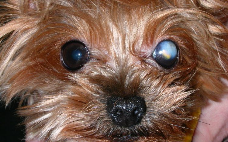
Not every cloudy eye in a dog indicates a cataract. Such changes can be caused by glaucoma, cataracts, uveitis, salt deposits in the eye tissue, or tumor tissue replacement.
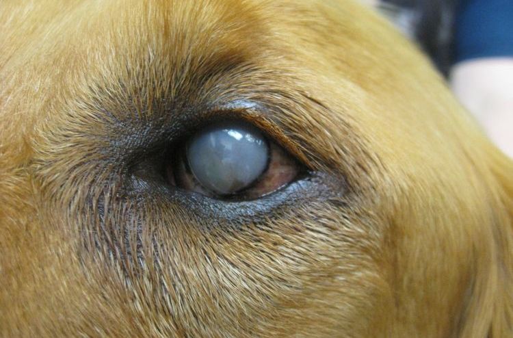
A cataract appears as a white spot, a cloudy cloud, or a widespread whitening, while the eye in the affected area loses its characteristic glossy, moist shine. It's important to note that the cataract itself doesn't cause pain or itching. Most often, a blind spot develops on the affected side of the eye: the dog becomes less responsive to stimuli, doesn't notice obstacles, and may become anxious if people or other animals suddenly appear in the area.
Reasons
- Age-related changes cause degeneration of corneal tissue. As a result of metabolic disturbances, cataract or nuclear sclerosis of the eye.
- Consequences hepatitis or a viral infection with signs of intoxication of the animal's body. In the initial stage, this may be keratitisThis disease is the second most common cause of eye cataracts in dogs, after age-related changes.
- Eye injury (chemical or mechanical). Appeared the ulcer leads to corneal degeneration.
- Erosions caused by bacterial infection of the eye.
- Consequences of eye surgery or as a result of neoplasms on the visual organ.
- Increased intraocular pressure leading to nerve death.
- Genetic predisposition. Typically, a dog's eye cataract in this case is the result of diabetes.
- Inversion of the eyelids, when the eyelashes injure the cornea.
- Congenital abnormalities. If the eyelids do not close tightly, the surface of the eye becomes dry.
Types of disease
- Peripheral. The edge of the eyeball becomes cloudy. Visually, you can see a whitish edge. Timely treatment stops the process, and visual acuity remains intact. A dog can live with this type of cataract for its entire life.
- Central. The center of the eye becomes cloudy. This is usually an age-related change that progresses over time. When completely blocked, the animal only reacts to light rays and cannot see objects.
- Total, when the eye is completely covered by a film. The dog suddenly loses sight and goes blind.
Symptoms
- Exudate discharge and frequent conjunctivitis. Increased discharge from the eyes after an injury should be especially alarming.
- A collection of pus in the corners of the eye. This is a sign of a bacterial infection of the visual organ. The cornea gradually becomes drier and rougher.
- Corneal clouding. The owner may notice a small whitish spot on their pet's eye that resolves within 1-2 days. This is superficial keratitis, a sign that the dog's vision needs to be checked. A short-term corneal clouding may be the result of an injury or a sign of hepatitis.
- Changes in corneal color. It may simply become cloudy or acquire a yellowish-reddish tint.
- Photophobia. The animal will be reluctant to go for walks during the day and will seek out darkened corners of the house.
- Deteriorating vision. This is evident in the dog's behavior: it becomes less confident in its actions, tries to stay close to its owner, and walks awkwardly. One sign of vision impairment is squinting.
The danger of disease
The appearance of a cataract in a dog's eye can lead not only to decreased or complete loss of vision, but also to complications of the underlying conditions that caused the clouding, discomfort, pain, and even the risk of losing the entire eye.
Trauma and inflammation that cause a cataract often affect not only the superficial cornea but also deeper structures of the eye—the iris, lens, and other tissues. Combined with a bacterial or fungal infection, this can lead to infection of the entire eye and require drastic measures, including removal of the eyeball. Long-term untreated chronic infections increase the risk of sepsis and the spread of microbes to other tissues and organs, not just the head.
Diagnostics
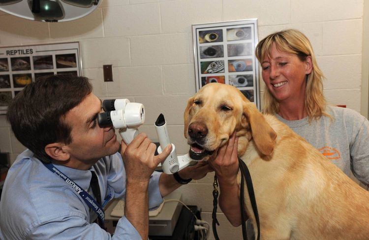
Before treating a cataract, the doctor conducts a full examination of the dog, which includes:
- Identification herpes virus.
- Corneal swabs to determine whether the disease is caused by a virus or bacteria.
- Corneal scraping. A traumatic procedure used when other diagnostic methods have been inconclusive. Its purpose is to obtain biopsy material to determine the type of infection.
- Examination of ocular fluid (paracentesis).
- Blood test if fungal infection is suspected.
- Examination of the retina using special ophthalmological instruments.
- Ultrasound of the eyeball. This method allows for examination in cases of total damage.
Treatment
The most common cause of the disease is a bacterial infection. After taking a swab and identifying the pathogen, the doctor will select an antibiotic and develop a treatment plan. Medications are used in the early stages of the disease and after surgery. There are standard treatment regimens:
- Removing exudate. Simply put, you need to regularly flush your dog's eyes. You can use regular boiled water or chamomile tea, but miramistin or furacilin are better options.
- Applying tetracycline ointment under the eyelid is the medication most often prescribed by veterinarians.
- Catarrhal lesions are treated with chloramphenicol ointment or solution, as well as hydrocortisone suspension.
- To prevent the spread of infection, Tobrex drops are instilled into the eye.
- If degenerative changes occur in the eye, you can't treat it on your own. You'll need the help of a veterinarian, who will prescribe injections.
Please note: Surgery is performed when medication alone fails to prevent the progression of pathological changes.
Preventive measures
- Regular check-ups by a veterinarian, deworming and vaccination.
- Daily hygiene procedures: examination of the visual organs, rinsing and wiping the eyes
- Avoiding eye injuries. Dogs can be injured by branches, sharp edges, or fighting.
- Self-medication is unacceptable. Only a specialist knows which remedies will not harm the eye or worsen the condition. Many folk remedies are acid-based, which can cause corneal burns.
Read also:


Add a comment